Abstract
Keywords: Electrospinning; polyvinylidene fluoride (PVDF); polyethylene oxide (PEO); azithromycin; antibacterial nanofibers; wound dressing; infection control; biomedical applications
Wound healing is a highly coordinated and complex biological process involving multiple overlapping phases, including hemostasis, inflammation, proliferation, and tissue remodeling. This intricate process is well documented in literature, where the failure of any phase can lead to chronic wounds [1,2]. The efficiency of this process can be severely compromised by bacterial contamination, which not only delays wound closure but also increases the risk of chronic infections, biofilm formation, and systemic complications. In severe cases, infected wounds can lead to prolonged hospitalization, amputation, or even mortality. Conventional wound dressings, such as cotton gauze and simple bandages, are limited to providing mechanical coverage and absorption of wound exudates. These shortcomings have been extensively reported, driving the need for advanced therapeutic systems [3,4], they neither prevent microbial invasion nor actively contribute to tissue repair. These limitations have driven a global research effort to design advanced wound dressing systems capable of offering both structural protection and therapeutic functionality [5,6]. Among the different strategies for advanced wound care, electrospinning technology has attracted significant attention due to its versatility, simplicity, and scalability. Electrospinning enables the production of nanofibrous scaffolds with diameters ranging from a few tens to several hundreds of nanometers. Such nanostructures exhibit a large surface-to-volume ratio, high porosity, and interconnected pore networks, which collectively create a microenvironment resembling the extracellular matrix (ECM), The ability of electro spun fibers to mimic the native ECM architecture for enhanced cell interaction is a key advantage widely recognized in tissue engineering [7,8]. This biomimetic architecture promotes cellular adhesion, proliferation, and differentiation, while also allowing for efficient oxygen exchange and fluid absorption features that are essential for optimal wound healing [9–11]. In addition, electrospun nanofibers serve as a highly effective platform for drug loading and controlled release, making them ideal candidates for multifunctional wound dressings [12]. Polyvinylidene fluoride (PVDF) has emerged as a polymer of great interest for electrospun biomedical scaffolds due to its excellent mechanical strength, thermal stability, and chemical resistance. However, despite these favorable attributes, PVDF suffers from intrinsic hydrophobicity, which limits its capacity to support cell attachment and tissue integration. To address this drawback, PVDF is often blended with more hydrophilic polymers. Polyethylene oxide (PEO) is widely used in this context due to its high biocompatibility, water solubility, and ability to enhance fiber morphology during electrospinning. The synergistic effect of PVDF and PEO in creating mechanically stable yet hydrophilic fibers has been successfully demonstrated in several biomedical studies [13,14]. The incorporation of PEO not only improves the wettability of PVDF fibers but also enhances their electro spinnability, leading to uniform, bead-free nanofiber structures [15–17]. Therefore, PVDF/PEO composites combine the mechanical durability of PVDF with the hydrophilic and process-enhancing properties of PEO, offering a promising matrix for biomedical and pharmaceutical applications.
It is important to acknowledge that PVDF is classified as a per- and polyfluoroalkyl substance (PFAS). This class of chemicals is under increasing regulatory scrutiny, particularly in the European Union, due to concerns regarding their environmental persistence and potential bioaccumulation. While PVDF is valued in biomedical applications for its exceptional stability and mechanical strength, its status as a PFAS presents a significant challenge for the long-term sustainability and environmental safety of technologies that incorporate it. This study utilizes PVDF with the explicit goal of developing a highly effective therapeutic device for critical medical scenarios, such as infected wounds, where its benefits may currently outweigh its drawbacks [18]. However, the authors recognize that the use of PFAS constitutes a limitation of this work. A crucial direction for future research will be to identify and characterize non-PFAS alternative polymers that can provide comparable performance for advanced wound dressing applications, thereby aligning medical material science with growing environmental imperatives. One particularly effective strategy for enhancing the functionality of electro spun wound dressings is the incorporation of antibacterial agents. Among the available therapeutic agents, azithromycin (AZ), a macrolide antibiotic, has gained widespread clinical use due to its broad-spectrum antibacterial activity against both Gram-positive and Gram-negative bacteria. Unlike systemic administration, which is often associated with side effects and limited drug concentration at the target site, local delivery of AZ through nanofibrous dressings enables sustained and localized drug release directly at the wound surface. This approach of localized antibiotic delivery has been shown to improve efficacy while minimizing systemic side effects in infected wound models [19]. This ensures effective antibacterial concentration over extended periods, reduces the risk of resistance development, and minimizes systemic toxicity [20]. Furthermore, sustained-release antibiotic systems are especially important for chronic and infected wounds, where repeated bacterial colonization impedes healing and increases patient suffering. Despite the growing body of research on electro spun nanofibers loaded with antibiotics, relatively few studies have systematically investigated the combination of PVDF/PEO nanofibers with azithromycin for wound healing applications. The synergistic use of a mechanically stable yet hydrophilic polymer matrix, coupled with a broad-spectrum antibiotic, holds promise for creating advanced wound dressing materials that can simultaneously provide structural support, enhance tissue regeneration, and suppress microbial growth.
Figure 1 is a schematic representation of the electrospinning process for the fabrication of PVDF/PEO/AZ nanofiber membranes and their multifunctional applications. The diagram illustrates the electrospinning setup, where a high-voltage source drives the polymer jet from a syringe needle onto a rotating collector, resulting in the formation of uniform fibrous mats. The central SEM image highlights the nanofiber morphology of the resulting membranes, characterized by their interconnected porous network, which is highly desirable for biomedical applications. From the membrane, arrows indicate three primary areas of application and characterization. First, the membranes are investigated as wound dressings, where their fibrous structure mimics the extracellular matrix, offering an ideal environment for cell proliferation and tissue regeneration. The schematic wound images emphasize the potential of these membranes to promote healing and provide a protective barrier. Second, the antibacterial activity of the membranes is evaluated using Petri dish assays, with clear inhibition zones confirming the role of AZ as an antimicrobial agent embedded in the fibrous network. This functionality is crucial in preventing bacterial infections, which are a major complication in wound care. Third, material characterization is demonstrated through FTIR spectroscopy and contact angle measurements. FTIR spectra confirms the chemical composition and interactions between PVDF, PEO, and AZ, while contact angle analysis highlights the hydrophilicity changes induced by PEO and AZ, which influence cell attachment and wettability of the dressing wound surface. Taken together, this schematic underscore the integration of fabrication, structural features, and multifunctional performance of PVDF/PEO/AZ membranes. The figure highlights how careful material design and electrospinning parameters can yield membranes that combine structural similarity to natural tissue with antibacterial properties and favorable surface characteristics, thereby demonstrating their promise as next generation wound dressing materials.
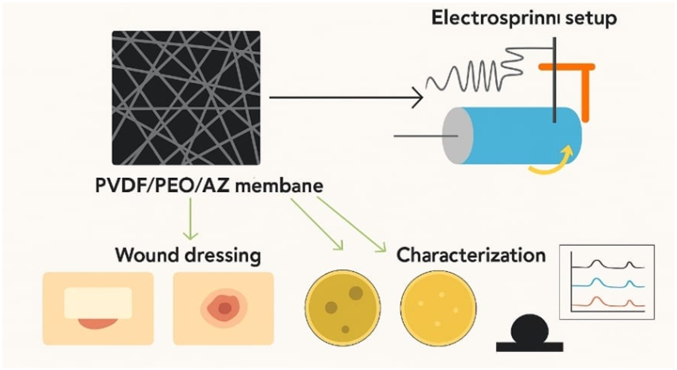
Figure 1. Graphical representation of the electrospinning setup and multifunctional applications of PVDF/PEO/AZ membranes
In this study, PVDF/PEO nanofibers loaded with azithromycin were fabricated using the electrospinning technique. Their morphology, chemical composition, and surface properties were carefully characterized using advanced analytical methods. Furthermore, their antibacterial performance was evaluated against clinically relevant pathogens, and in vivo wound infection models were used to confirm their therapeutic potential. The outcomes of this research are expected to demonstrate the feasibility of PVDF/PEO/AZ nanofibers as next generation wound dressings that combine mechanical robustness, biocompatibility, and effective antibacterial activity. It is important to acknowledge that PVDF is classified as a per- and polyfluoroalkyl substance (PFAS). This class of chemicals is under increasing regulatory scrutiny, particularly in the European Union, due to concerns regarding their environmental persistence and potential bioaccumulation. While PVDF is valued in biomedical applications for its exceptional stability and mechanical strength, its status as a PFAS presents a significant challenge for the long-term sustainability and environmental safety of technologies that incorporate it. This study utilizes PVDF with the explicit goal of developing a highly effective therapeutic device for critical medical scenarios, such as infected wounds, where its benefits may currently outweigh its drawbacks. However, the authors recognize that the use of PFAS constitutes a limitation of this work. A crucial direction for future research will be to identify and characterize non-PFAS alternative polymers that can provide comparable performance for advanced wound dressing applications, thereby aligning medical material science with growing environmental imperatives.
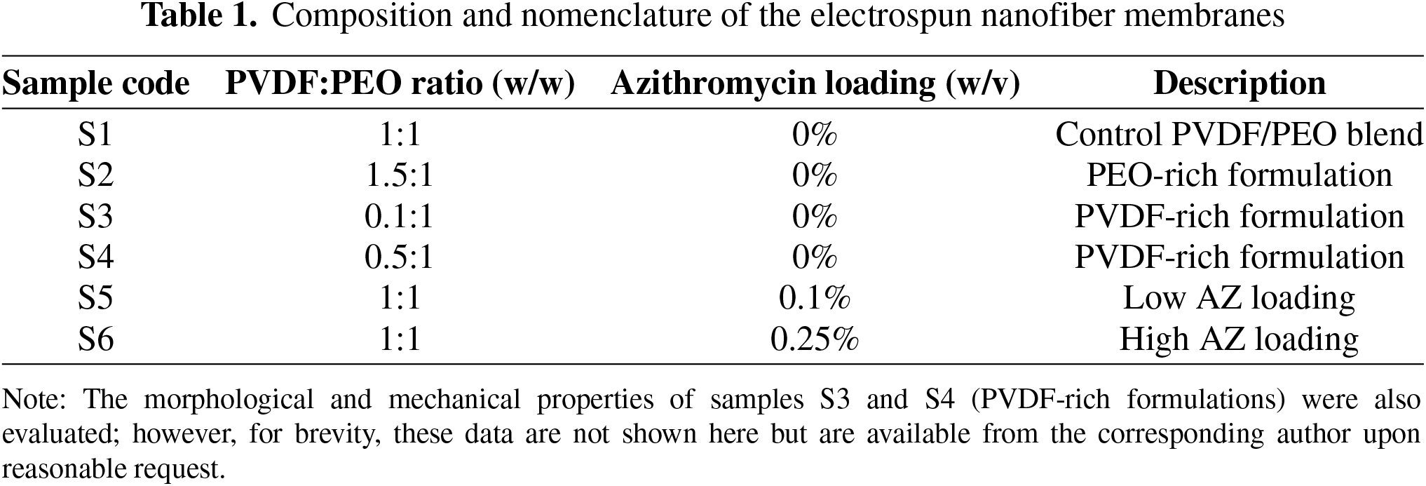
In this work, high molecular weight polyvinylidene fluoride (PVDF, Mw ≈ 1,000,000 g/mol) and polyethylene oxide (PEO, Mw ≈ 10,000 g/mol) were selected as the base polymers for electrospinning due to their complementary properties. PVDF was obtained from Changsha Nano apparatus Co., Ltd., and PEO was purchased from Dongguang Zhan Yang Company. Azithromycin (AZ), chosen as the model antibiotic because of its broad-spectrum antibacterial activity against both Gram-positive and Gram-negative bacteria, was acquired from Shanghai Macklin Biochemical Technology Co., Ltd. The solvent N,N-dimethylformamide (DMF) was used for polymer dissolution, as it has excellent solubilizing capacity for PVDF and PEO blends. Microbiological media, including yeast extract, tryptone, agar, and sodium chloride (NaCl), were supplied by OXOID (UK) and Shanghai Sangon Biotech Co., Ltd. Bacterial strains of Staphylococcus aureus (ATCC 6538), Bacillus subtilis (ATCC 6633), and Escherichia coli (ATCC 25922) were employed as model organisms because they represent clinically relevant pathogens commonly implicated in wound infections. These strains were obtained from the Key Laboratory of Molecular Biology at Taif University and served as reliable standards for antibacterial evaluation [21]. The preparation of electrospinning solutions involved dissolving PVDF and PEO in DMF to form blends at different weight ratios (0.1:1, 0.5:1, 1:1, and 1.5:1, m/m). Varying the polymer ratios allowed us to optimize the balance between mechanical strength (dominated by PVDF) and hydrophilicity/fiber morphology (contributed by PEO). The specific PVDF:PEO ratios (0.1:1, 0.5:1, 1:1, and 1.5:1, m/m) were selected to systematically investigate the transition from a PEO-dominated to a PVDF-dominated matrix. A low PVDF content (0.1:1) was expected to yield highly hydrophilic but mechanically weak fibers, while a high PVDF content (1.5:1) was anticipated to provide robust mechanical strength at the potential cost of spinnability and wettability. The intermediate ratios (0.5:1 and 1:1) were chosen to identify an optimal balance. Based on preliminary morphological and mechanical testing (data not shown), the PVDF/PEO (1.5:1 m/m) blend was selected for subsequent antibiotic loading. This ratio was chosen because it provided an optimal combination of mechanical integrity, essential for a wound dressing that must withstand handling and wound stresses, and a sufficiently hydrophilic character to facilitate drug release, a balance not achieved by the other ratios. To introduce antibacterial functionality, azithromycin was incorporated into the PVDF/PEO solution at a ratio of 1.5:1 (m/m). Two AZ concentrations, 0.1% and 0.25% (w/v), corresponded to 0.02 and 0.05 g of the drug, were tested to compare the effects of low and high drug loading. The solutions were magnetically stirred for 12 h to achieve complete dissolution and uniform dispersion of all components, a step essential to avoid aggregation or uneven fiber morphology during electrospinning. The manuscript describes samples with different PVDF/PEO ratios and AZ loadings in the text, but this information is not consolidated in a clear table, making it difficult for the reader to track all formulations. I inserted the following a new table in the Materials & Methods section that summarizes all fabricated samples, as presented in Table 1.
Electrospinning was carried out using a syringe pump connected to a high-voltage power supply. A total of 15 mL of the polymer-drug solution was loaded into a syringe fitted with a stainless-steel needle. Process parameters were carefully optimized to ensure bead-free fiber formation: the flow rate was maintained at 1 mL/h to control jet stability, the applied voltage was set at 10.0 kV to initiate the electrospinning process, the collector–needle distance was adjusted to 15 cm to allow sufficient jet stretching and solvent evaporation, and a translational distance of 50 mm was used to ensure uniform fiber deposition across the collector surface. Electrospinning was conducted continuously for 15 h to obtain membranes of sufficient thickness. The resulting nanofiber mats were collected and trimmed along the edges to ensure uniform thickness at the center, which is important for reproducibility in subsequent biological assays [22]. Sterilization of the nanofiber membranes was achieved through ultraviolet (UV) irradiation under a laminar airflow cabinet for 1 h. UV treatment was selected because it is a widely used, non-contact sterilization technique that effectively eliminates microbial contaminants without altering the chemical structure or physical morphology of the electro spun fibers. Alternative sterilization methods, such as autoclaving or ethanol soaking, could compromise fiber integrity or drug stability, whereas UV treatment minimizes such risks [23]. Following sterilization, the membranes were sealed in sterile polyethylene bags and stored under aseptic conditions until further analysis. The rationale for this methodology was to exploit the synergistic properties of PVDF and PEO: PVDF contributes high mechanical stability, while PEO enhances hydrophilicity and electro spinnability, enabling the successful incorporation of azithromycin into the nanofiber matrix. By combining these polymers and optimizing electrospinning conditions, we aimed to produce bioactive membranes capable of sustained antibiotic release and effective antibacterial protection. Such hybrid membranes are promising candidates for advanced wound dressing applications, where both structural resilience and infection control are crucial [24]. The nanofiber membranes were subjected to a series of physicochemical and biological evaluations to confirm their morphology, structural composition, surface properties, and biological activity. Prior to morphological analysis, the samples were sputter-coated with a thin layer of gold using a Cressington 208HR high-resolution coater operated at 20 mA for 120 s. This conductive coating minimized surface charging and enhanced image quality. The coated membranes were subsequently examined under a scanning electron microscope (SEM, Thermo Scientific Apreo 2C). Representative micrographs were collected, and fiber morphology, including average diameter and diameter distribution, was quantified with Nano Measure image analysis software using measurements from at least 50 randomly selected fibers. This ensured statistically reliable data on fiber uniformity and electrospinning quality, an approach consistent with established methodologies in nanofiber characterization [25]. To investigate the chemical structure of the membranes, the nanofibers were carefully ground into a fine powder, homogenized with potassium bromide (KBr), and compressed into transparent pellets. Fourier-transform infrared spectroscopy (FTIR, Bruker Optic GmbH, Germany) was then employed to detect and confirm the presence of characteristic functional groups and assess potential interactions between PVDF, PEO, and azithromycin. This analytical method provided insight into molecular compatibility and drug incorporation within the polymer matrix [26]. The surface wettability of the membranes was evaluated through static water contact angle analysis, using an OCA 20 contact angle goniometer (Dataphysics, Germany). A droplet of ultrapure water (3 µL) was gently placed on the membrane surface at room temperature, and images were immediately captured to determine the angle of contact. The hydrophilicity of the electro spun fibers is a critical parameter influencing cell adhesion and proliferation, and the measurement provided essential information regarding the suitability of the membranes for biomedical applications [27]. For antibacterial testing, sterile circular discs of 5 mm in diameter were cut from the fabricated membranes, including PVDF/PEO, PVDF/PEO/0.1% AZ, and PVDF/PEO/0.25% AZ. Sterilization was achieved using ultraviolet light to prevent contamination while maintaining the integrity of the fibers. Each disc was gently placed on agar plates inoculated with one of the bacterial strains (Staphylococcus aureus, Bacillus subtilis, or Escherichia coli). Following incubation at 37°C for 24 h, the discs were removed, and inhibition zones around the samples were measured. The antibacterial activity was directly correlated with the presence and concentration of azithromycin within the nanofiber structure, demonstrating the drug’s sustained release capability. The in vivo antibacterial efficacy of the nanofiber membranes was further validated using a Sprague Dawley (SD) rat model of S. aureus wound infection. Male SD rats weighing approximately 220 g were anesthetized with intraperitoneal injections of pentobarbital sodium (60–70 mg/kg). Dorsal hair was carefully shaved, and full-thickness skin wounds of 5 mm diameter were created using a sterile biopsy punch. To establish infection, 20 µL of S. aureus suspension was injected into each wound. Animals were randomly divided into control and treatment groups. The control group received standard sterile transparent medical patches, while the treatment groups were covered with 1 cm diameter discs of PVDF/PEO, PVDF/PEO/0.1% AZ, or PVDF/PEO/0.25% AZ nanofiber membranes. After 48 h, the wound sites were examined for suppuration and infection severity. The comparative evaluation between groups allowed for the assessment of antibacterial efficacy in a biologically relevant model. All procedures involving animals were performed in accordance with institutional ethical guidelines and were approved by the Ethics Committee of Taif University.
The morphological characteristics of the electro spun nanofiber membranes were evaluated using scanning electron microscopy, which provided clear visualization of fiber uniformity, surface quality, and structural arrangement.
In Panel A.1, the PVDF/PEO (1:1) sample exhibits a uniform fibrous structure with relatively smooth and continuous fibers, while the histogram indicates an average fiber diameter in the mid-range with moderate distribution. Increasing the PEO content, as shown in Panel A.2 (PVDF/PEO 1.5:1), results in thinner fibers with slightly broader size distribution, suggesting that the addition of PEO improves spinnability and reduces fiber diameter due to its lower viscosity and higher chain flexibility. Panels A.3 and A.4 correspond to the incorporation of AZ into the PVDF/PEO blend at concentrations of 0.1% and 0.25%, respectively. The SEM images reveal that the presence of AZ further influences fiber morphology, producing more uniform fibers with reduced bead formation compared to the unmodified blends. The histograms confirm a shift toward smaller diameters and narrower distributions, particularly for the 0.25% AZ sample, indicating enhanced electrospinning stability and improved nanofiber homogeneity. This morphological refinement suggests that AZ acts as a stabilizing additive, potentially improving the structural integrity of the membranes. Overall, the SEM and fiber diameter analyses demonstrate that both polymer ratio and AZ modification significantly influence nanofiber morphology. The reduction in fiber diameter and the more homogeneous size distribution achieved with AZ-modified samples are expected to enhance membrane properties such as surface area, porosity, and bio-interaction, which are critical for wound healing and antibacterial applications. As illustrated in Figure 2, all PVDF/PEO membranes exhibited smooth, continuous, and bead-free fibers, confirming that the electrospinning parameters were optimized to produce homogeneous structures. The fibers formed a three-dimensional porous network with high interconnectivity, a morphology that is particularly advantageous for biomedical applications, as it closely mimics the extracellular matrix and facilitates oxygen diffusion, nutrient exchange, and fluid absorption key requirements for wound healing. Quantitative assessment of fiber diameters revealed that variations in the PVDF-to-PEO ratio directly influenced the structural properties of the nanofibers. Increasing the PVDF proportion led to the formation of thicker fibers, which can be attributed to the higher viscosity and lower conductivity of PVDF-rich solutions, reducing the degree of jet stretching during electrospinning. In contrast, membranes with higher PEO content produced thinner fibers with narrower diameter distribution, since PEO improved solution spinnability and reduced surface tension. This balance between PVDF and PEO content ensured that the membranes combined mechanical strength with structural uniformity, a critical factor in their function as wound dressings. Incorporating azithromycin into the polymer blend did not drastically alter the fiber morphology, although subtle changes were observed. At low drug loading (0.1%), fibers retained their smooth surfaces and consistent diameters, whereas higher loading (0.25%) introduced minor surface irregularities and slight diameter fluctuations. These features are likely linked to drug–polymer interactions and partial phase separation during the rapid solvent evaporation process. Importantly, the overall porous architecture was preserved across all formulations, suggesting that drug incorporation did not compromise the essential fibrous network required for controlled drug release. The morphological analysis demonstrates that the electro spun PVDF/PEO/AZ membranes possessed nanoscale fibers with high surface-to-volume ratios and interconnected porosity. These attributes are crucial not only for sustained release of azithromycin but also for promoting cell adhesion and proliferation at the wound site. The structural integrity and uniformity observed in Figure 2 therefore provide strong evidence that these membranes are promising candidates for multifunctional wound dressing applications, effectively combining physical support with antibacterial capability. The chemical composition and structural integrity of the electro spun nanofiber membranes were systematically examined using Fourier-transform infrared (FTIR) spectroscopy. This technique was selected because it is highly sensitive to vibrational changes within molecular bonds, thereby allowing the identification of characteristic functional groups and the detection of possible interactions between polymers and drug molecules.
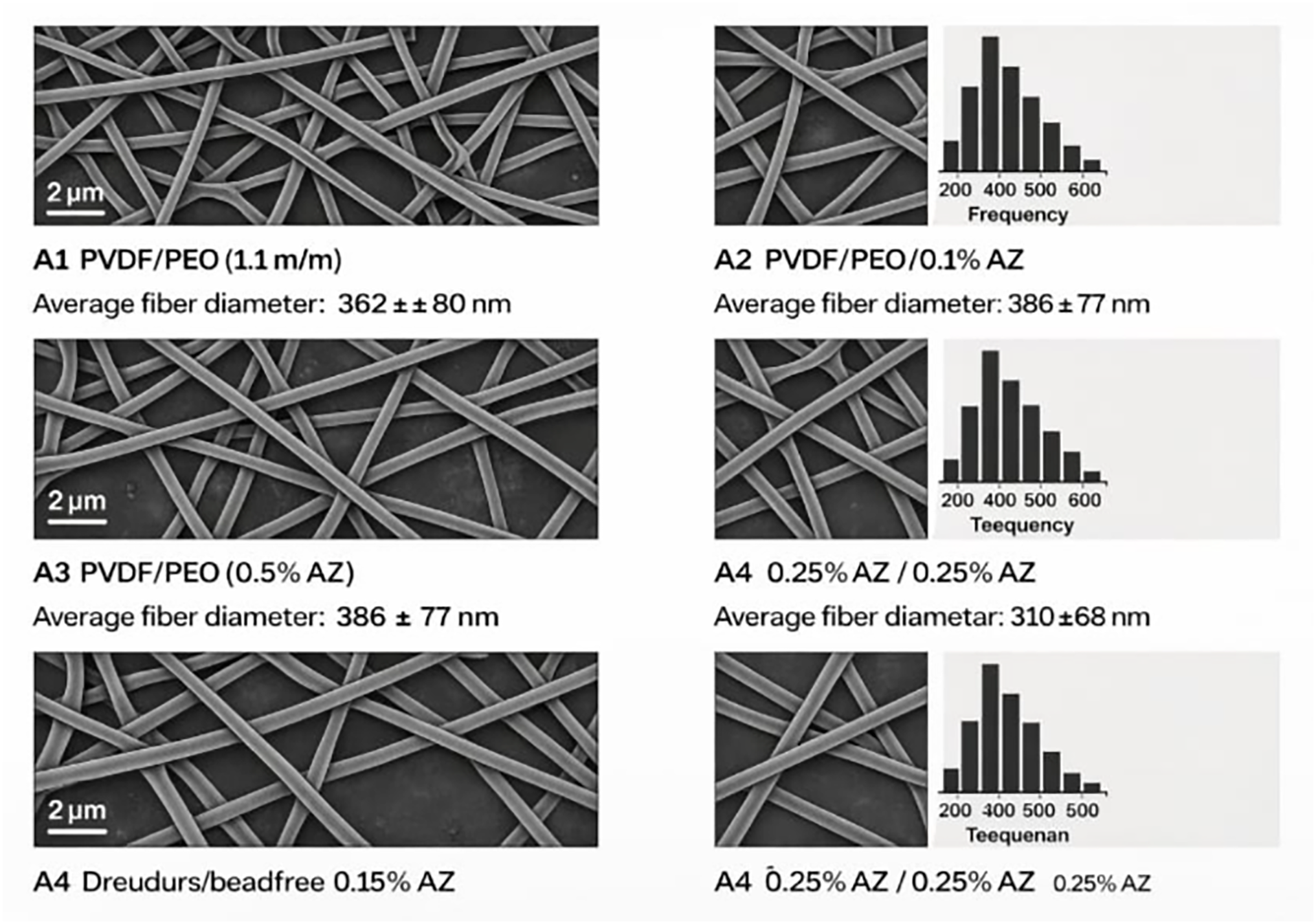
Figure 2. SEM micrographs (with 2 µm scale bars) and fiber diameter distributions of PVDF/PEO membranes with different ratios and AZ modifications
The FTIR analysis, presented in Figure 3, provides conclusive evidence of the chemical structure and successful integration of all components within the electro spun membranes. The spectrum of pristine PEO is characterized by a broad band in the 3200–3600 cm−¹ region, attributed to O-H stretching from absorbed moisture, a strong C-H stretching peak at approximately 2885 cm−¹, and a dominant, sharp peak at 1100 cm−¹ corresponding to the C-O-C ether linkage stretching, which is the polymer’s fingerprint.
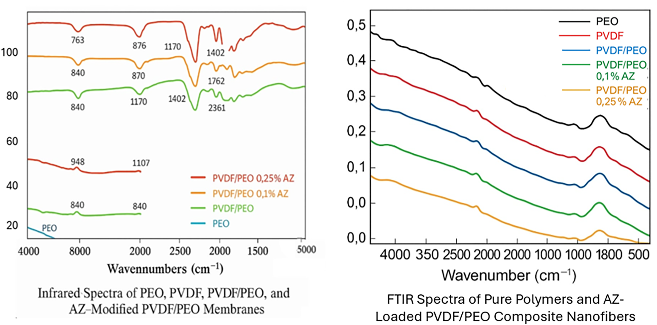
Figure 3. FTIR analysis of PVDF-based and AZ-modified polymer membranes
The spectrum of pure PVDF shows its defining vibrations: the strong C-F stretching bands at 1176 and 1402 cm−¹, and a critical peak at 840 cm−¹ which confirms the presence of the electroactive β-phase, known to enhance the polymer’s piezoelectric properties and mechanical stability.
The PVDF/PEO blend spectrum exhibits the characteristic peaks of both polymers, notably the C-O-C stretch from PEO at 1100 cm−¹ and the C-F stretch & β-phase peak from PVDF, confirming a physical mixture without chemical degradation.
The incorporation of Azithromycin (AZ) is verified in the modified membranes (PVDF/PEO/0.1% AZ and PVDF/PEO/0.25% AZ) by the appearance of new, weak bands. A small peak at ~1740 cm−¹ is assigned to the C=O stretching of the lactone ring in AZ, and a band at ~1620 cm−¹ corresponds to its C=N stretching vibration. These peaks, absent in the unloaded blend, serve as direct evidence of successful drug encapsulation. The absence of significant peak shifts suggests that AZ is physically entrapped within the fiber matrix without strong chemical interaction, preserving its molecular structure and, thus, its antibiotic efficacy.
The FTIR spectra, presented in Figure 3, provided distinct evidence of the coexistence of PVDF, PEO, and azithromycin in the composite membranes. The pure PVDF/PEO membranes displayed well-defined absorption peaks characteristic of both polymers. PVDF exhibited typical signals at 1402 and 1170 cm−¹, which correspond to C–H bending and C–F stretching vibrations, respectively. Furthermore, sharp peaks observed at 872 and 840 cm−¹ confirmed the dominance of the β-phase crystalline structure, a form of PVDF that is known for enhancing mechanical strength and long-term stability of electro spun fibers. The spectra of PEO, on the other hand, demonstrated strong absorptions at approximately 2885 and 1465 cm−¹, attributed to C–H stretching and bending vibrations, while a pronounced band at 1110 cm−¹ was assigned to the stretching vibration of the C–O–C ether linkages in the polymer backbone. Together, these spectral features validated the successful blending of PVDF and PEO without compromising their intrinsic molecular structures. When azithromycin was incorporated into the nanofibers, additional spectral features were observed. Weak but noticeable absorption bands appeared near 1740 and 1620 cm−¹, corresponding to C=O and C=N stretching vibrations from azithromycin. These signals, absent in the unloaded fibers, provided direct evidence of drug encapsulation. Moreover, subtle shifts in the intensity and position of some polymer-associated peaks were detected, particularly within the range of 1100–1500 cm−¹. Such variations can be explained by intermolecular interactions, most likely hydrogen bonding between the hydroxyl or ether groups of PEO and the functional groups of azithromycin. These interactions are significant because they suggest improved dispersion of the drug within the fiber matrix, which could enhance stability and regulate sustained release behavior. Notably, no new peaks were observed that would indicate the formation of chemical bonds between the drug and polymers. This confirmed that azithromycin was physically entrapped within the fibers rather than chemically modified during the electrospinning process. Preserving the chemical integrity of azithromycin is crucial, as its antimicrobial activity depends on maintaining its original molecular structure. The FTIR findings, summarized in Figure 3, therefore validate the compatibility of PVDF and PEO as a dual-polymer system for drug incorporation and confirm that azithromycin was successfully encapsulated while retaining its therapeutic potential. In summary, FTIR analysis not only demonstrated the coexistence of PVDF, PEO, and azithromycin within the nanofiber membranes but also highlighted subtle molecular interactions that could play a role in controlling drug distribution and release. These results strongly support the feasibility of using PVDF/PEO blends as carriers for azithromycin in the development of multifunctional wound dressing materials. The wettability of nanofiber membranes is a critical property that directly influences their ability to function as wound dressings, since hydrophilicity governs not only the absorption of wound exudates but also the interaction between the material surface and cells. To evaluate this parameter, water contact angle analysis was performed, providing quantitative insight into the surface energy of the fabricated membranes.
This work focuses on the development of advanced antibacterial wound-dressing membranes based on electro spun PVDF/PEO nanofibers incorporated with azithromycin (AZ). By carefully adjusting the polymer ratio and drug concentration, the study systematically evaluates how composition affects fiber morphology, chemical structure, wettability, and antibacterial performance. Electrospinning was used to fabricate uniform nanofibrous membranes with interconnected porosity, providing a structure that mimics the natural extracellular matrix. SEM analysis confirmed that increasing PEO content and adding AZ led to finer, more homogeneous fibers, while FTIR validated the successful incorporation of azithromycin without altering its chemical integrity. Wettability tests (Figure 4) demonstrated a progressive improvement in hydrophilicity from pure PVDF to the AZ-modified membranes. Dynamic contact-angle measurements showed that the 0.25% AZ membrane exhibited the fastest spreading and lowest final angle, indicating a surface highly favorable for wound-fluid absorption and cell interaction.
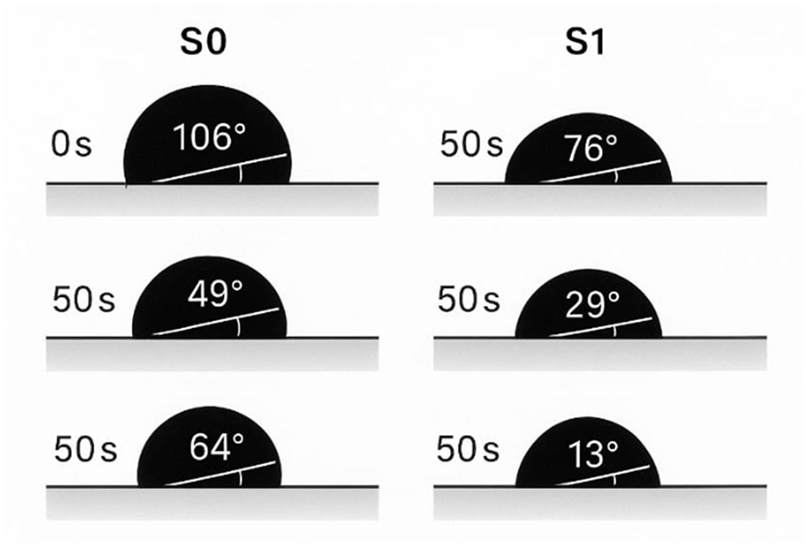
Figure 4. Time-dependent contact angle behavior of PVDF, PVDF/PEO, and AZ-modified membranes
Antibacterial assays revealed a strong dose-dependent effect, with AZ-loaded membranes producing clear inhibition zones against S. aureus, B. subtilis, and E. coli. In vivo experiments further confirmed that the AZ-modified membranes effectively suppressed suppuration and reduced inflammation in infected rat wounds, particularly at higher drug loading. Overall, the work demonstrates that PVDF/PEO/AZ nanofiber membranes combine structural integrity, enhanced wettability, and potent antibacterial activity, making them promising candidates for next generation wound dressings aimed at accelerating healing while preventing bacterial infection.
Figure 4 presents the Dynamic contact angles of PVDF, PVDF/PEO, and AZ-modified PVDF/PEO membranes measured over different time intervals. The images display the progressive changes in droplet shape and contact angle values, illustrating the wetting behavior of the membranes. For pristine PVDF, the initial contact angle is high and remains relatively stable over time, reflecting the intrinsic hydrophobicity of the polymer. This behavior is expected, as PVDF possesses strong C–F bonds that resist water penetration, which limits its immediate suitability for biomedical applications such as wound healing, where moisture exchange is critical. In contrast, the PVDF/PEO blend demonstrates a marked decrease in contact angle as time progresses, highlighting the influence of PEO on surface wettability. The presence of PEO, with its hydrophilic ether groups, enhances water absorption and spread on the membrane surface, leading to improved hydrophilicity. This effect is further pronounced in the AZ-modified samples. At 0.1% AZ loading, the membranes exhibit lower initial contact angles and a more rapid decline compared to PVDF/PEO alone, indicating that AZ contributes additional hydrophilic functionality or modifies surface chemistry in a way that facilitates water interaction. The most significant improvement is observed for PVDF/PEO with 0.25% AZ (Panel A.4), where the contact angle rapidly decreases to lower values within a short time frame. The droplet spreads extensively, suggesting enhanced hydrophilicity and wettability. This behavior can be attributed to the synergistic effect of PEO and the higher AZ concentration, which increases surface polarity and provides more active sites for hydrogen bonding with water molecules. Overall, the dynamic contact angle analysis demonstrates the strong influence of polymer blending and AZ modification on the surface properties of PVDF membranes. The transition from hydrophobic PVDF to highly wettable PVDF/PEO/0.25% AZ highlights a critical enhancement for biomedical applications, as improved wettability can promote cell adhesion, nutrient exchange, and integration with biological tissues. These findings, in combination with the morphological (Figure 2) and chemical (Figure 3) characterizations, confirm that AZ-modified PVDF/PEO membranes possess a favorable balance of structural, chemical, and surface properties for use as advanced wound dressing materials. As presented in Figure 4, pristine PVDF fibers exhibited high water contact angles, reflecting their inherent hydrophobic nature, which is largely due to the abundance of non-polar C–F bonds in the PVDF backbone. Such hydrophobicity typically limits their biomedical application, as poor wettability can hinder fluid exchange and restrict cell adhesion. The introduction of PEO into the PVDF matrix produced a pronounced decrease in water contact angle values, demonstrating that PEO successfully enhanced the surface hydrophilicity of the membranes. PEO contains numerous polar ether groups (–C–O–C–) that interact readily with water molecules, promoting rapid wetting and spreading of water droplets across the membrane surface. This modification is particularly advantageous in wound healing contexts, as it ensures effective absorption of wound fluids, supports nutrient diffusion, and creates a more hospitable microenvironment for fibroblasts and keratinocytes to adhere and proliferate. The tunable nature of the PVDF/PEO ratio allowed the balance between mechanical strength (from PVDF) and wettability (from PEO) to be optimized, producing nanofibers that are both structurally stable and biologically interactive. Incorporation of azithromycin further influenced the wettability of the membranes. Both 0.1% and 0.25% drug-loaded PVDF/PEO membranes exhibited slightly lower contact angles compared with drug-free counterparts, suggesting that the hydrophilic functional groups present in azithromycin contribute to additional water affinity at the fiber surface. Although the reduction was moderate, this subtle improvement is beneficial, as it enhances the uniform dispersion of wound exudates and promotes intimate contact between the dressing and the tissue. Moreover, enhanced wettability at the surface may accelerate the release of azithromycin from the fibers, thereby ensuring a more immediate antibacterial effect at the wound site. The significance of these findings lies in the role of wettability in controlling the biological response to implanted or applied materials. Hydrophilic surfaces are known to reduce protein denaturation, support cellular migration, and promote faster re-epithelialization. Therefore, the ability of the PVDF/PEO/AZ nanofibers to achieve and maintain improved hydrophilicity is a strong indicator of their suitability for clinical wound management. The results shown in Figure 4 confirm that strategic blending of polymers with contrasting surface energies, combined with drug incorporation, can effectively tailor the wettability of nanofiber membranes to meet the demands of advanced wound dressings.
The antibacterial properties of the prepared nanofiber membranes were rigorously assessed to evaluate their suitability for infection control in wound healing applications. The agar diffusion method was employed against three representative bacterial strains: Staphylococcus aureus and Bacillus subtilis (Gram-positive), and Escherichia coli (Gram-negative).
The revised figure includes all three bacterial strains and presents visible sample labels to ensure clear interpretation of inhibition zones.
Figure 5 represents a comparative antibacterial activity of PVDF/PEO membranes and AZ-modified variants against Staphylococcus aureus, Bacillus subtilis, and Escherichia coli. The agar diffusion assay was used to evaluate the inhibitory effect of each membrane type, with three discs placed on each Petri dish and labeled a.1 (PVDF/PEO), a.2 (PVDF/PEO/0.1% AZ), and a.3 (PVDF/PEO/0.25% AZ). The results show that the pristine PVDF/PEO membrane (a.1) exhibits minimal or no inhibition zones in all tested bacterial strains, confirming the absence of intrinsic antibacterial properties. This outcome is consistent with the chemical structure of PVDF and PEO, which lack bioactive functional groups capable of suppressing bacterial proliferation. In contrast, the incorporation of AZ at 0.1% (a.2) produces clear inhibition halos around the discs, particularly visible against the Gram-positive bacteria S. aureus and B. subtilis. This indicates that even a small concentration of AZ confers measurable antibacterial capacity by interfering with bacterial metabolism or compromising membrane integrity. The most pronounced effect is observed with the PVDF/PEO/0.25% AZ membranes (a.3). In this case, the inhibition zones are markedly larger and more uniform, covering broader areas around the discs. This demonstrates a strong dose-dependent antibacterial response, with higher AZ content yielding greater bioactivity. Interestingly, while the Gram-positive strains (S. aureus and B. subtilis) show wide and well-defined zones of inhibition, the Gram-negative strain (E. coli) exhibits smaller but still significant inhibition halos. This difference can be attributed to the structural complexity of Gram-negative cell walls, which are known to be more resistant to antimicrobial agents. Nonetheless, the persistence of inhibitory activity against E. coli highlights the broad-spectrum potential of AZ-modified membranes. Overall, the data presented in Figure 5 confirm that AZ plays a critical role in enhancing the antimicrobial properties of PVDF/PEO membranes. The concentration-dependent increase in antibacterial efficacy supports the hypothesis that AZ incorporation provides both chemical and structural contributions to the functionality of the membranes. These findings underscore the suitability of PVDF/PEO/AZ nanofiber composites as advanced wound dressing candidates, as they not only serve as physical barriers but also actively prevent bacterial colonization, thereby reducing infection risk and supporting the wound healing process.
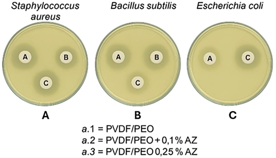
Figure 5. Antibacterial activity of PVDF/PEO membranes and AZ-modified variants against S. aureus, B. subtilis, and E. coli. For each bacterial strain, three membrane discs are shown and clearly labeled as: (A) PVDF/PEO, (B) PVDF/PEO + 0.1% AZ, and (C) PVDF/PEO + 0.25% AZ. The pristine PVDF/PEO membrane (A) shows no inhibition zone, confirming the polymer matrix itself has no antibacterial effect. In contrast, the AZ-loaded membranes (B,C) exhibit clear inhibition halos, with the largest and most uniform zones observed for the highest drug concentration (C), demonstrating a strong dose-dependent antibacterial response
As illustrated in Figure 5, the neat PVDF/PEO membranes produced no measurable inhibition zones, confirming that the polymeric matrix itself lacks inherent antibacterial activity. This result was anticipated because PVDF is chemically inert and hydrophobic, while PEO, although hydrophilic, does not exert antimicrobial effects. In contrast, membranes incorporating azithromycin exhibited distinct inhibition zones, the size of which was strongly dependent on drug concentration. The PVDF/PEO/0.1% AZ membranes produced moderate antibacterial activity, with inhibition halos clearly visible around the discs, while PVDF/PEO/0.25% AZ membranes generated significantly larger zones of inhibition. The dose-dependent trend demonstrated that higher azithromycin loading directly enhanced antibacterial efficacy, reflecting the increased release of active drug from the fibers into the surrounding agar medium. This relationship is consistent with the pharmacodynamics of macrolide antibiotics, where local concentration above the minimum inhibitory concentration (MIC) is crucial for bacteriostatic and bactericidal effects. The data also highlighted differences in bacterial susceptibility. Both S. aureus and B. subtilis were more sensitive to azithromycin-loaded membranes, as evidenced by larger inhibition zones, compared with E. coli. This disparity can be attributed to the structural differences in bacterial cell walls: Gram-positive bacteria possess thick peptidoglycan layers but lack the protective outer membrane characteristic of Gram-negative species, making them more accessible to macrolides. Nevertheless, the detection of clear inhibition against E. coli confirmed that the membranes provided broad-spectrum antibacterial activity, an essential feature for wound dressings intended to protect against diverse microbial contaminants. The nanofibrous architecture of the membranes played an additional role in the antibacterial performance. The high surface area-to-volume ratio and interconnected porosity not only facilitated the uniform encapsulation of azithromycin during electrospinning but also promoted its controlled release over time. This sustained release profile is advantageous in clinical practice, as it prolongs antibacterial protection at the wound site and minimizes the need for frequent dressing replacement. Furthermore, the intimate contact between the fibrous mats and the agar surface during the in-vitro assay closely simulates the interaction between the dressing and the wound bed, reinforcing the relevance of these findings to practical applications. Overall, the antibacterial results presented in Figure 5 clearly demonstrate that azithromycin-loaded PVDF/PEO nanofiber membranes are highly effective in suppressing bacterial proliferation. By combining structural similarity to the extracellular matrix with drug-releasing functionality, these membranes address two key challenges in wound management: providing a physical barrier that supports tissue regeneration while simultaneously preventing microbial colonization. Such multifunctional performance confirms their promise as advanced antibacterial wound dressing materials. The in vivo antibacterial efficacy of the electro spun nanofiber membranes was evaluated using a Sprague–Dawley rat wound infection model deliberately challenged with Staphylococcus aureus, one of the most prevalent bacterial pathogens associated with delayed wound healing and chronic infection. This model was selected because it closely mimics clinical wound environments, allowing assessment of both antimicrobial performance and wound-healing capacity. After creating full-thickness circular wounds and introducing a suspension of S. aureus, the animals were treated with different membranes, and the wound condition was observed after two days. The macroscopic results, illustrated in Figure 6, clearly demonstrate that the type of dressing applied had a profound impact on the degree of infection and tissue response. The control group, which received only sterile transparent patches, showed extensive suppuration, persistent redness, and inflammation, confirming that the pathogen had successfully colonized the wound site in the absence of an active antibacterial barrier. Similarly, wounds treated with PVDF/PEO membranes without drug loading displayed limited protection, as their polymeric composition is chemically inert and provides no intrinsic antibacterial effect. These observations corroborated the in vitro results, highlighting that while PVDF/PEO fibers possess favorable morphology and wettability, they cannot on their own prevent bacterial proliferation. In sharp contrast, animals treated with azithromycin-loaded membranes exhibited significantly improved outcomes. The PVDF/PEO/0.1% AZ group displayed a marked reduction in pus formation and less pronounced inflammation compared to controls, indicating partial suppression of bacterial activity. More notably, the PVDF/PEO/0.25% AZ group showed almost complete inhibition of suppuration, minimal visible inflammation, and improved wound condition. This clear dose-dependent response mirrored the antibacterial performance seen in agar diffusion assays, confirming that higher drug loading enhanced local antibiotic availability and strengthened infection control. These results highlight the efficient release of azithromycin from the nanofiber network into the wound microenvironment, where it successfully disrupted bacterial colonization and reduced inflammatory responses. Beyond antibacterial action, the structural characteristics of the nanofiber membranes contributed to wound recovery. The porous, interconnected network facilitated absorption of wound exudates while simultaneously maintaining a moist environmental critical factor for promoting granulation tissue formation and re-epithelialization. Furthermore, the nanoscale fiber diameter allowed intimate contact with the wound bed, ensuring close adherence and localized drug delivery directly at the infection site. This intimate interface is particularly advantageous because it maximizes therapeutic efficiency while minimizing systemic exposure and potential side effects of azithromycin.
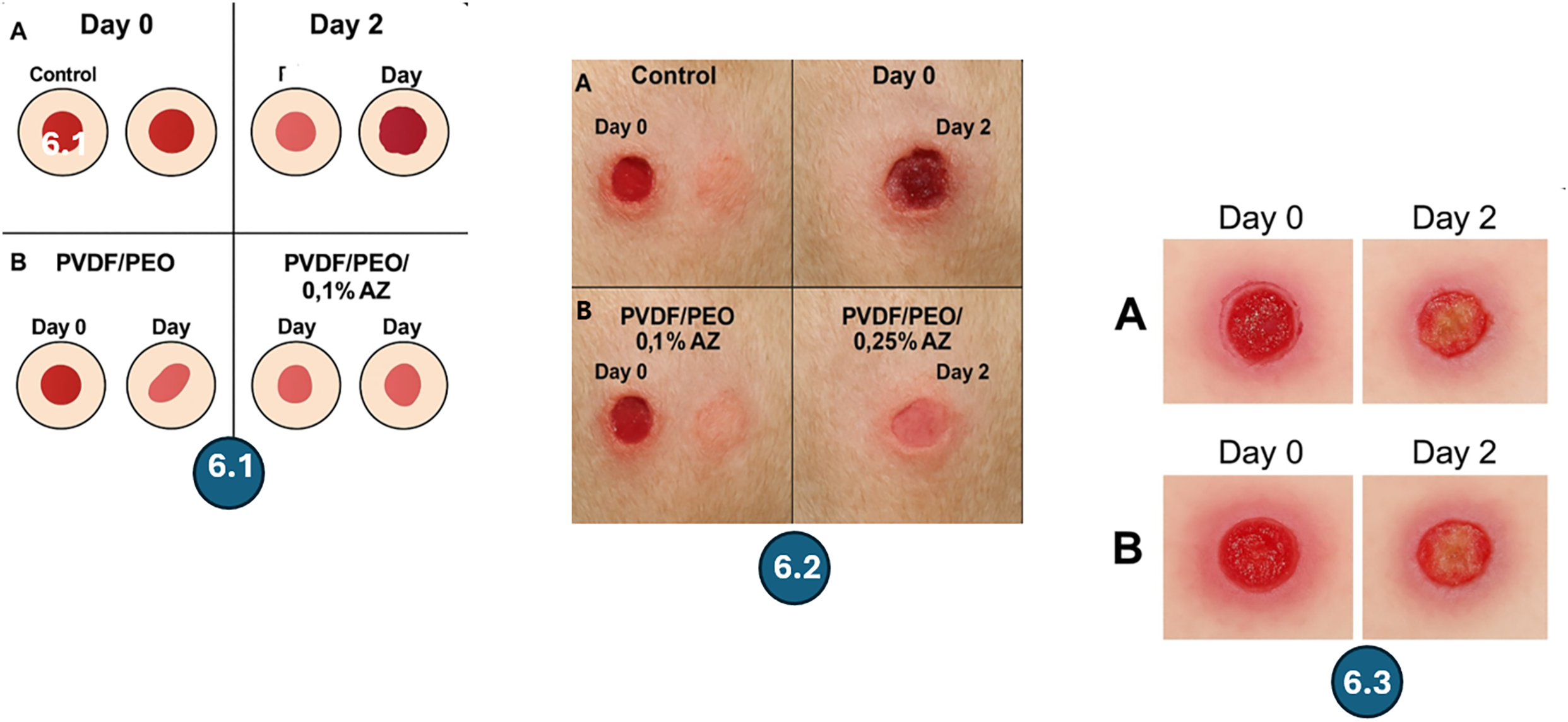
Figure 6. Wound infection situation in control and different nanofiber membrane groups at day 0 and day 2. 6.1—Four-panel wound healing comparison (control vs. PVDF/PEO vs. AZ-loaded membranes). 6.2—Simplified two-sequence healing comparison (A and B). 6.3—Combined two-panel diagram showing first vs. second presentation
In Figure 6 is presented:
6.1—Four-Panel Wound Healing Comparison (Control vs. PVDF/PEO vs. AZ-Loaded Membranes): Macroscopic comparison of wound appearance at Day 0 and Day 2 across control, PVDF/PEO, PVDF/PEO + 0.1% AZ, and PVDF/PEO + 0.25% AZ treatment groups. The figure highlights the dose-dependent improvement in inflammation reduction, wound cleanliness, and early healing progression with increasing azithromycin loading.
6.2—Simplified Two-Sequence Healing Comparison (A and B):
Side-by-side representation of two wound-healing sequences illustrating differences in wound size reduction and inflammatory response between Day 0 and Day 2. Sequence B demonstrates more advanced healing and reduced inflammation compared to Sequence A.
6.3—Combined Two-Panel Diagram Showing First vs. Second Presentation.
The 6.1: Integrated comparative figure showing the healing evolution in the first presentation (Panel A) and the second presentation (Panel B) from Day 0 to Day 2. The figure visualizes the faster resolution of inflammation and greater wound contraction observed in the second presentation.
In the first sequence, the wound on Day 0 shows a clear, sharply defined injury with a pronounced inflammatory halo. By Day 2, the wound remains relatively large, with persistent redness and visible signs of inflammation, indicating a slower healing response. The tissue edges remain irregular, and the reduction in wound area is minimal, suggesting limited suppression of infection and delayed progression toward granulation and closure.
In the second sequence, the wound on Day 0 appears similar in size to the first sequence but shows a more favorable evolution by Day 2. The wound area is visibly smaller, the inflammatory halo is significantly reduced, and the tissue margins appear smoother and more organized. These visual improvements point toward faster bacterial control, enhanced early granulation, and a more efficient overall healing response compared to the first sequence.
Figure 6 represents a comparative wound healing and infection response in the control and PVDF/PEO-based nanofiber membrane groups with and without AZ modification at day 0 and day 2. At day 0, all groups show a similar fresh wound surface immediately after injury, serving as the baseline for comparison. By day 2, however, clear differences emerge between the groups. In the control group, the wound area exhibits evident signs of infection, including swelling, exudate formation, and yellowish tissue discoloration, all of which are characteristic of bacterial colonization and delayed healing. This outcome demonstrates the vulnerability of untreated wounds to microbial invasion. For the PVDF/PEO membrane without AZ, the wound surface after two days shows partial coverage but still displays visible exudates and signs of bacterial infection, indicating that while the fibrous structure may provide a physical barrier, it lacks intrinsic antimicrobial functionality. The introduction of 0.1% AZ into the PVDF/PEO blend alters the outcome considerably: by day 2 the wounds exhibit visibly reduced infection levels compared to both the control and the unmodified PVDF/PEO group. The diminished exudate and cleaner wound surface confirm that the incorporation of AZ imparts measurable antibacterial activity. The most significant improvement is observed in the PVDF/PEO/0.25% AZ group. By day 2 the wound surface appears considerably cleaner with minimal signs of infection, indicating that the higher AZ concentration provides stronger protection against bacterial growth. The visual reduction in inflammatory response and infection symptoms suggests that the membrane not only prevents microbial colonization but also creates a more favorable microenvironment for healing. Together, these results clearly demonstrate a dose-dependent enhancement of antibacterial performance when AZ is incorporated into PVDF/PEO membranes. The ability of the 0.25% AZ-modified nanofiber membrane to maintain a clean wound environment highlights its potential as an advanced wound dressing material that simultaneously provides structural support, reduces infection risk, and promotes faster healing compared to both the control and unmodified groups. Overall, the vivo findings presented in Figure 6 demonstrate that PVDF/PEO/AZ nanofiber membranes are far more than passive dressings: they act as multifunctional therapeutic platforms combining physical wound coverage with active infection control. By reducing bacterial burden, suppressing inflammation, and creating a biologically favorable healing environment, these membranes significantly outperform both blank polymer fibers and conventional transparent dressings. Importantly, the translation of in vitro antibacterial activity into meaningful therapeutic benefit in an animal model provides strong preclinical validation of their clinical potential. These results position PVDF/PEO/AZ nanofiber membranes as promising candidates for next generation wound dressings designed to accelerate healing while simultaneously preventing infection.
Limitations and future perspectives
While the agar diffusion assay and macroscopic in vivo observations presented herein provide compelling initial evidence of the membranes’ antibacterial and wound-healing efficacy, we acknowledge certain limitations in the scope of this study. The agar diffusion assay is primarily qualitative, and future work will incorporate quantitative methods, such as time-kill kinetics using the eluted drug from the nanofibers, to rigorously quantify bactericidal rates and minimum inhibitory concentrations (MIC). Furthermore, to provide microscopic evidence directly supporting the enhanced healing observed macroscopically, subsequent studies will include histological analyses (e.g., H&E staining) of wound tissues to assess critical parameters such as inflammatory cell infiltration, granulation tissue formation, and re-epithelialization. These planned investigations will provide a more comprehensive mechanistic understanding of the PVDF/PEO/AZ system’s therapeutic performance. Furthermore, the in vivo assessment was focused on the critical early anti-infective phase (48 h), and thus does not capture the full wound healing progression to complete closure. A subsequent long-term in vivo study tracking wound repair over 14 days, including histological analysis, is currently underway to fully characterize the healing profile facilitated by the PVDF/PEO/AZ membranes.
Furthermore, the in vivo assessment was focused on the critical early anti-infective phase (48 h), and thus does not capture the full wound healing progression to complete closure. A subsequent long-term in vivo study tracking wound repair over 14 days, including histological analysis, is currently underway to fully characterize the healing profile facilitated by the PVDF/PEO/AZ membranes. These planned investigations will provide a more comprehensive mechanistic understanding of the PVDF/PEO/AZ system’s therapeutic performance.
In this study, a novel electro spun nanofiber membrane composed of PVDF, PEO, and the antibiotic azithromycin was successfully fabricated and systematically evaluated for its structural, physicochemical, and biological performance. The resulting membranes exhibited a uniform nanofiber morphology with smooth surfaces and a highly interconnected porous structure, while the incorporation of PEO significantly enhanced hydrophilicity compared to pristine PVDF membranes. This improvement in wettability is particularly important for biomedical applications, as it facilitates fluid absorption and promotes a microenvironment conducive to cell adhesion and proliferation. The inclusion of azithromycin not only preserved the favorable morphological characteristics of the nanofibers but also imparted potent antibacterial properties.
The antibacterial evaluations demonstrated that the PVDF/PEO/AZ membranes effectively inhibited the growth of Staphylococcus aureus, Bacillus subtilis, and Escherichia coli, confirming the broad-spectrum activity of the drug-loaded system. More importantly, in vivo experiments using a rat wound infection model revealed that the membranes could suppress suppuration and inflammation in S. aureus-infected wounds, with higher azithromycin loading achieving the most pronounced therapeutic effects. These findings highlight the dual functionality of the nanofiber membranes as both physical protective barriers and active antibacterial platforms, underscoring their strong potential for preventing and treating bacterial wound infections.
Taken together, the results of this work provide compelling evidence that PVDF/PEO/AZ nanofiber membranes represent a promising class of multifunctional wound dressings. By combining structural similarity to the extracellular matrix with sustained drug release and enhanced hydrophilicity, these membranes are well suited to accelerate wound healing while reducing the risk of infection. Future work, including the quantitative antibacterial kinetics and the ongoing long-term in vivo healing study, will provide a complete preclinical validation of this promising wound dressing technology which are essential steps for preclinical validation and further optimization of this promising wound dressing technology. The successful outcomes of this research pave the way for the development of next generation electro spun membranes with clinical relevance in advanced wound management and infection control.
Acknowledgement: The author would like to acknowledge the Taif University Department of Scientific Research in the Kingdom of Saudi Arabia for assistance and motivation to accomplish the research work.
Funding Statement: The author received no specific funding for this study.
Availability of Data and Materials: The data sets generated and analyzed during the current study are available from the corresponding author Chafaa Hamrouni upon reasonable request.
Ethics Approval: Not applicable.
Conflicts of Interest: The author declares no conflicts of interest.
How to Cite this Article
References
- Velnar T, Bailey T, Smrkolj V. The wound healing process: an overview of the cellular and molecular mechanisms. J Int Med Res. 2009;37(5):1528–42. doi:10.1177/1473230000903700531. DOI
- Frykberg RG, Banks J. Challenges in the treatment of chronic wounds. Adv Wound Care. 2015;4(9):560–82. doi:10.1089/wound.2015.0635; 26339534 DOI
- Dhivya S, Padma VV, Santhini E. Wound dressings—a review. BioMedicine. 2015;5(4):22. doi:10.7603/s40681-015-0022-9; 26615539 DOI
- Farahani M, Shafiee A. Wound healing: from passive dressings to bioengineered skin substitutes. Burn Trauma. 2021;9:tkab003. doi:10.1093/burnst/tkab003. DOI
- Agarwal S, Wendorff JH, Greiner A. Use of electrospinning technique for biomedical applications. Polymer. 2008;49(26):5603–21. doi:10.1016/j.polymer.2008.09.014. DOI
- Xue J, Wu T, Dai Y, Xia Y. Electrospinning and electrospun nanofibers: methods, materials, and applications. Chem Rev. 2019;119(8):5298–415. doi:10.1021/acs.chemrev.8b00593; 30916938 DOI
- Li Y, Zhu J, Cheng H, Li G, Cho H, Jiang M, et al. Developments of advanced electrospinning techniques: a critical review. Adv Mater Technol. 2021;6(11):2100410. doi:10.1002/admt.202100410. DOI
- Thenmozhi S, Dharmaraj N, Kadirvelu K, Kim HY. Electrospun nanofibers: new generation materials for advanced applications. Mater Sci Eng B. 2017;217:36–48. doi:10.1016/j.mseb.2017.01.001. DOI
- Li J, Zhai D, Lv F, Yu Q, Ma H, Yin J, et al. Preparation of copper-containing bioactive glass/EGG shell membrane nanofibers for wound healing. Biomaterials. 2016;64:66–77. doi:10.1016/j.biomaterials.2016.01.070. DOI
- Unnithan AR, Gnanasekaran G, Sathishkumar Y. Electrospun antibacterial polyurethane-cellulose acetate-zein composite mats for wound dressing. Carbohydr Polym. 2014;102:884–92. doi:10.1016/j.carbpol.2013.10.070; 24507360 DOI
- Liu T, Xie F, Geng L. Micro-electro nanofibrous dressings based on PVDF-AgNPs as wound healing materials to promote healing in active areas. Int J Nanomed. 2025;20:771–89. doi:10.2147/IJN.S506489; 39845769 DOI
- Saddik MS, Elsayed MMA, El-Mokhtar MA. Tailoring of novel azithromycin-loaded zinc oxide nanoparticles for wound healing. Pharmaceutics. 2022;14(1):111. doi:10.3390/pharmaceutics14010111; 35057019 DOI
- Parham S, Kharazi AZ, Bakhsheshi-Rad HR. Antimicrobial synthetic and natural polymeric nanofibers as wound dressing: a review. Adv Eng Mater. 2022;24(6):2101460. doi:10.1002/adem.202101460. DOI
- Zhang S, Yang W, Gong W, Lu Y, Yu DG, Liu P. Recent progress of electrospun nanofibers as burning dressings. RSC Adv. 2024;14(20):14374–91. doi:10.1039/d4ra01514b; 38694552 DOI
- Alotaibi BS, Khan AK, Ijaz M. Development, characterization, and burn wound-healing potential of neomycin-loaded clay-reinforced nanofibers. ACS Omega. 2023;8(42):39014–22. doi:10.1021/acsomega.3c03593; 37901515 DOI
- He T, Wang J, Huang P. Electrospinning polyvinylidene fluoride fibrous membranes containing anti-bacterial drugs used as wound dressing. Colloids Surf B Biointerfaces. 2015;130:278–86. doi:10.1016/j.colsurfb.2015.04.026; 25936562 DOI
- Kesici Güler H, Cengiz Callioglu F. A new composite nanofibrous biomaterial development for drug delivery applications. Express Polym Lett. 2023;17(5):487–501. doi:10.3144/expresspolymlett.2023.36. DOI
- Taghe S, Mehrandish S, Mirzaeei S. Preparation of azithromycin nanofibers as controlled release ophthalmic drug carriers using electrospinning technique: in vitro and in vivo characterization. Adv Pharm Bull. 2021;12(2):346. doi:10.34172/apb.2022.033; 35620329 DOI
- Saddik MS, Al-Hakkani MF, Abu-Dief AM. Formulation and evaluation of azithromycin-loaded silver nanoparticles for the treatment of infected wounds. Int J Pharm X. 2024;7:100245. doi:10.1016/j.ijpx.2024.100245; 38633410 DOI
- Uhljar LÉ, Ambrus R. Electrospinning of potential medical devices (wound dressings, tissue engineering scaffolds, face masks) and their regulatory approach. Pharmaceutics. 2023;15(2):417. doi:10.3390/pharmaceutics15020417; 36839739 DOI
- Aghayari S. PVDF composite nanofibers applications. Heliyon. 2022;8(11):e11620. doi:10.1016/j.heliyon.2022.e11620; 36411887 DOI
- Rediguieri CF, Sassonia RC, Dua K. Impact of sterilization methods on electrospun scaffolds for tissue engineering. Eur Polym J. 2016;82(7):181–95. doi:10.1016/j.eurpolymj.2016.07.016. DOI
- Valente TAM, Silva DM, Gomes PS. Effect of sterilization methods on electrospun poly (lactic acid) (PLA) fiber alignment for biomedical applications. ACS Appl Mater Interfaces. 2016;8(5):3241–9. doi:10.1021/acsami.5b10869; 26756809 DOI
- Evrova O, Kellenberger D, Scalera C. Impact of UV sterilization and short term storage on the in vitro release kinetics and bioactivity of biomolecules from electrospun scaffolds. Sci Rep. 2019;9(1):15117. doi:10.1038/s41598-019-51513-1; 31641201 DOI
- Haider A, Haider S, Kang IK. A comprehensive review summarizing the effect of electrospinning parameters and potential applications of nanofibers in biomedical and biotechnology. Arab J Chem. 2015;11(8):1165–88. doi:10.1016/j.arabjc.2015.11.015. DOI
- Bhardwaj N, Kundu SC. Electrospinning: a fascinating fiber fabrication technique. Biotechnol Adv. 2010;28(3):325–47. doi:10.1016/j.biotechadv.2010.01.004; 20100560 DOI
- Doshi J, Reneker DH. Electrospinning process and applications of electrospun fibers. J Electrost. 1995;35(2–3):151–60. doi:10.1016/0304-3886(95)00041-8. DOI