Abstract
Keywords: Adhesive agent; antioxidant; resveratrol; microtensile bond strength
In modern restorative dental practice, adhesive techniques form the basis of most minimally invasive procedures [1]. Adhesive systems must exhibit appropriate physicochemical properties together with favorable biological behavior to ensure predictable clinical performance [2]. Consequently, substantial research has focused on optimizing these characteristics to improve the longevity and reliability of adhesive restorations. A durable union between dentin and the restorative material is fundamental for the longevity and reliability of restorations [3]. Antioxidants incorporated into adhesive formulations have been shown to reduce hydrolytic and free-radical–mediated degradation processes [4], leading to extensive research into various antioxidant compounds. Among these, resveratrol (RES) has attracted attention due to its efficacy even at low concentrations [5].
RES is a naturally occurring polyphenol that has attracted considerable attention because of its potent antioxidant activity. It is predominantly found in grape-derived tissues and several plant-based food sources, where it functions as a secondary metabolite involved in defense mechanisms [6]. In addition to its powerful antioxidant properties, RES exhibits a wide range of biological effects. It is notable for its effects in controlling inflammation, preventing the development of cancer, protecting nerve tissue and slowing down the ageing process [7,8]. It also exhibits enhanced biocompatibility by mitigating oxidative stress, limiting the formation of free radicals, and protecting against DNA damage [9]. The addition of RES to adhesive formulations has been shown to markedly reduce cytotoxic effects and provide genoprotective benefits for fibroblasts, thereby enhancing the overall biocompatibility of these materials [9].
The durability of the resin–dentin interface is compromised when the depth of adhesive monomer penetration does not correspond to the level of collagen demineralization [10,11]. Under these conditions, collagen fibrils located at the base of the hybrid layer become susceptible to enzymatic degradation over time [12]. Such progressive breakdown of the hybrid layer promotes microleakage and nanoleakage, thereby increasing the risk of secondary caries and ultimately contributing to the clinical failure of the restoration [13]. RES has been reported to suppress the activity of matrix metalloproteinases (MMPs), particularly members of the MMP family such as MMP-2 and MMP-9 [14,15]. In addition, it inhibits glycolytic acid production and glucosyltransferase activity in Streptococcus mutans thereby providing potential protective effects against cariogenic biofilms [16].
The impact of RES on adhesive performance may vary depending on the formulation of the adhesive system. The antioxidant addition should enhance the biocompatibility of the material without compromising its mechanical or adhesive properties to ensure reliable clinical outcomes. Assessment of bonding effectiveness is commonly performed using tensile-based testing methods, which provide valuable information regarding the mechanical integrity and durability of adhesive interfaces [17].
Our previous investigation demonstrated that incorporating 0.5 μM RES into dentin adhesive systems did not compromise short-term μTBS values, indicating that RES may enhance the biological profile of adhesive formulations without adversely affecting their immediate adhesive performance [18]. However, short-term outcomes alone are insufficient to predict the clinical reliability of adhesive interfaces, as degradation processes primarily manifest over extended periods. Therefore, comprehensive long-term assessments are required to better approximate the material’s clinical behavior.
Although previous findings have demonstrated that RES-containing adhesive systems may improve dentin bond durability and exhibit antibiofilm potential [19], available evidence regarding their long-term adhesive performance after prolonged aging remains limited, particularly among different one-step self-etch adhesive formulations. Since the long-term stability of the adhesive interface is directly associated with restoration survival and secondary caries prevention [20], further long-term investigations are clinically relevant to determine whether RES incorporation can provide sustainable adhesive stability under extended aging conditions.
This study evaluated the impact of RES addition on the μTBS of five commercially available one-step self-etch adhesives after two years of water aging. It was hypothesized that the inclusion of RES would preserve the bonding performance of the adhesive systems and would not result in a significant reduction in bond strength following long-term storage.
2.1. Microtensile bond strength (μTBS) testing
The study was approved by the Ege University Human Ethics Committee (Approval No. 16-10.1/1), and written informed consent was obtained from all participants enrolled in the study prior to tooth extraction. This study utilized caries-free human third molars, which were first disinfected in 0.1% thymol solution and subsequently preserved in distilled water until the preparation of specimens. The roots were separated 2 mm apical to the cemento-enamel junction (CEJ), after which the occlusal enamel was removed 2 mm coronal to the CEJ using a low-speed, water-cooled diamond saw (Isomet; Buehler, Lake Bluff, IL, USA) to expose flat mid-coronal dentin. Following complete removal of the pulpal tissue, the dentin surfaces were finished with 600-grit silicon carbide (SiC) abrasive paper under continuous water irrigation for 60 s to create a standardized smear layer. The prepared teeth were randomly distributed into ten experimental groups (n = 6), in accordance with established protocols for microtensile bond strength testing [21,22].
Five one-step self-etch adhesives—G-aenial Bond (GC)/G, Optibond All-in-One (Kerr)/O, Gluma Self Etch (Kulzer)/Gl, Clearfil S3 Bond (Kuraray)/C, and Nova Compo-B Plus (Imicryl)/N—were evaluated with (+) and without (−) RES incorporation. Based on previous findings demonstrating enhanced fibroblast viability at 0.5 μM RES [9], this concentration was selected for the experimental groups. RES (Sigma-Aldrich, Saint Louis, USA) was added directly to the adhesive bottle immediately before application and homogenized by gentle agitation to prevent antioxidant degradation. According to the manufacturers’ guidelines, all adhesive materials were applied and subsequently photoactivated for 20 s using an LED curing unit (Elipar Freelight, 3M ESPE, St. Paul, MN, USA).
The composite resin (Filtek Z250; 3M ESPE, St. Paul, MN, USA) was incrementally placed onto each bonded surface in two 2-mm-thick layers. Each increment was light-cured for 20 s according to the manufacturer’s recommendations. Subsequently, the specimens were stored in distilled water at 37°C for a period of two years. Following the aging procedure, each tooth was sectioned perpendicular to the adhesive interface using a diamond saw to obtain beam-shaped specimens (1.00 ± 0.003 × 1.00 ± 0.003 mm2). Peripheral beams containing enamel were excluded; twenty central beams from each group were randomly selected for microtensile testing (n = 20).
The beams were attached to the testing jig with cyanoacrylate adhesive and subsequently tested in tension at a crosshead speed of 1.0 mm/min using a microtensile testing system (Microtensile Tester, BISCO Inc., Schaumburg, IL, USA). Following fracture, the exact cross-sectional dimensions of each specimen were determined with a digital caliper, and μTBS values were computed and expressed in MPa. Values obtained from beams originating from the same tooth were averaged and treated as a single experimental unit for statistical analysis, in accordance with μTBS testing guidelines. Fracture surfaces were evaluated with a stereomicroscope (LG-P52, Olympus, Tokyo, Japan) at ×50 magnification and categorized into adhesive, cohesive within dentin, cohesive within resin, or mixed failure types. The assessments and testing procedures were performed in a blinded fashion by another investigator using pre-assigned group codes. The design of the study is presented in Figure 1.
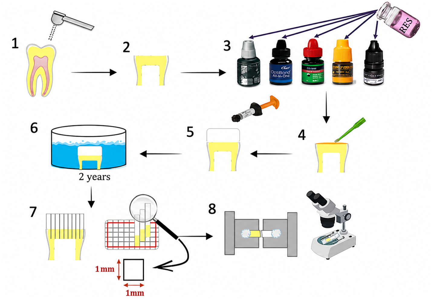
Figure 1. Workflow of RES-modified adhesives and long-term μTBS testing: (1,2) preparation of caries-free human third molar specimens; (3) preparation of five adhesive systems with or without 0.5 μM resveratrol (RES); (4) adhesive application and polymerization; (5) incremental composite resin build-up; (6) storage in distilled water for two years; (7) preparation of 1 × 1 mm2 beam specimens; and (8) microtensile bond strength testing followed by stereomicroscopic failure mode analysis.
Statistical analysis was carried out using GraphPad Prism software (version 5.0; GraphPad Software, La Jolla, CA, USA). Data are presented as mean ± standard deviation (SD). The Shapiro–Wilk test was used to assess normality, whereas Bartlett’s test was applied to evaluate the equality of variances. One-way analysis of variance (one-way ANOVA) was used to determine differences between groups. Pairwise comparisons among groups were subsequently carried out using Tukey’s post-hoc test whenever significant differences were present. The level of statistical significance was set at α = 0.05.
The mean μTBS values of all experimental groups are shown in Figure 2. After two years of water aging, adhesive-type failures predominated in all groups, whereas mixed failures were detected only in specimens from the O (+) and O (−) groups. No cohesive failures in dentin or composite were observed (Figure 3).
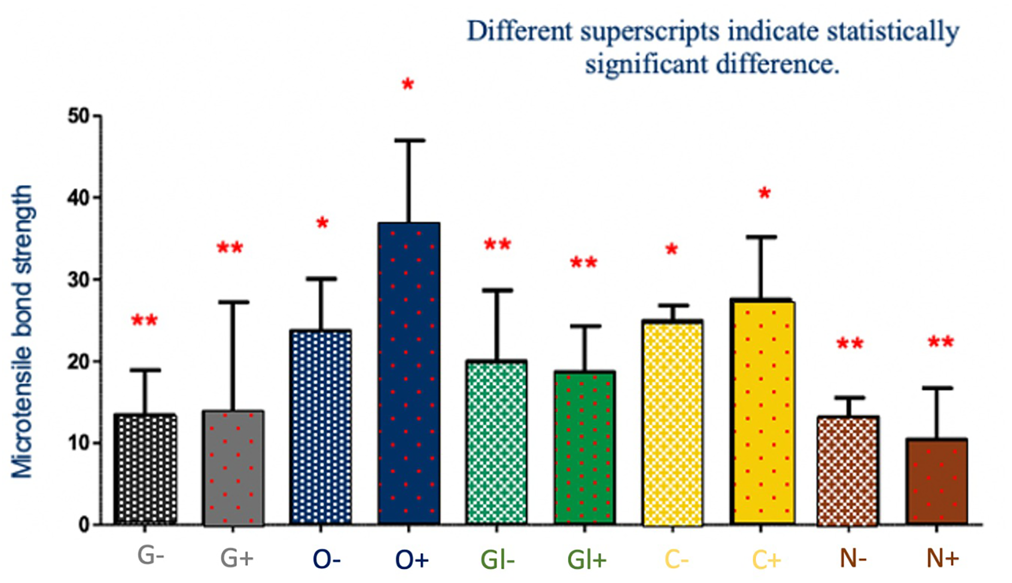
Figure 2. μTBS values of the adhesive systems with and without RES incorporation after two years of water aging. (G-aenial Bond (GC)/G, Optibond All-in-One (Kerr)/O, Gluma Self Etch (Kulzer)/Gl, Clearfil S3 Bond (Kuraray)/C, and Nova Compo-B Plus (Imicryl)/N, +: RES incorporation, −: without RES). (Different superscripts indicate statistically significant differences among the groups. Groups marked with the same symbol (* or **) were not significantly different from each other (p > 0.05), whereas groups marked with different symbols (* versus **) showed statistically significant differences (p < 0.05))
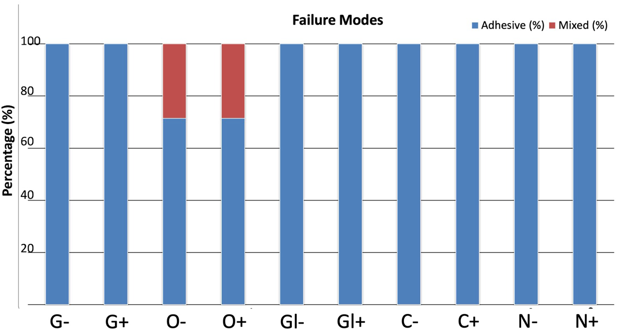
Figure 3. Failure modes of tested groups. (G-aenial Bond (GC)/G, Optibond All-in-One (Kerr)/O, Gluma Self Etch (Kulzer)/Gl, Clearfil S3 Bond (Kuraray)/C, and Nova Compo-B Plus (Imicryl)/N, +: RES incorporation, −: without RES)
RES incorporation did not result in a reduction in μTBS for any of the tested one-step self-etch adhesives when compared with their corresponding control groups. Among the materials evaluated, O (+) exhibited the highest mean bond strength, followed by C (+). Statistical analysis revealed no significant difference between O (+) and C (+) groups (p > 0.05).
For the control formulations (−), the μTBS values of G, Gl, and N groups were statistically similar (p > 0.05). This pattern was also maintained in the RES-modified versions of these adhesives, with no significant differences detected among their μTBS means (p > 0.05). Within each adhesive system, RES addition caused an increase in μTBS to varying degrees; however, for G, Gl, and N groups, this increase was not statistically significant.
Comparisons between adhesive systems showed that both O (+/−) and C (+/−) groups demonstrated significantly higher μTBS values than G, Gl, and N groups, regardless of RES incorporation (p < 0.05). Conversely, the three lower-performing adhesives (G, Gl, and N groups) did not differ significantly from one another (p > 0.05).
Dental materials are in constant contact with the oral environment; therefore, achieving optimal biocompatibility is a fundamental requirement in restorative dentistry. Although a completely biocompatible material is not yet achievable, alternative approaches have been explored to minimize the cytotoxic effects of resin monomers, and antioxidants have emerged as promising candidates in this regard [23]. Importantly, strategies aimed at improving the biological performance of adhesives must not compromise their adhesive capacity.
RES is a naturally occurring polyphenol abundant in grapes, berries and nuts, and is distinguished by its strong antioxidant properties [24,25]. In addition to its antioxidative effects, previous studies have demonstrated its antimicrobial potential. It has been reported that RES exhibits bacteriostatic effects against several Gram-positive species [26], while other investigations have shown that RES-rich Polygonum cuspidatum extracts suppress glycolytic acid formation, glucosyltransferase (GLT) activity [27], and various virulence factors of Streptococcus mutans [28]. These findings highlight RES as a multifunctional molecule capable of influencing both biological and microbiological pathways relevant to resin–dentin interface degradation.
Polyphenols promote collagen cross-linking and remineralization through their phenolic hydroxyl groups while also inhibiting MMP activity [29–31]. These phenolic hydroxyl groups contribute to the preservation of collagen integrity within the hybrid layer by suppressing MMP-mediated collagen degradation through multiple mechanisms [32]. RES, a naturally occurring polyphenol, may therefore reduce nanoleakage and improve dentin–resin bond strength when applied after contamination by preserving the structural integrity of the dentin–adhesive interface. This effect has been attributed particularly to the inhibition of MMP-9 activity, which enhances the resistance of dentin collagen to enzymatic degradation and promotes collagen cross-linking and stabilization [33].
The varying diffusion properties of adhesive monomers, which play a critical role in the adhesion process, may explain the differing interactions observed between RES and various monomer combinations. Incorporation of RES into adhesive systems has been reported to enhance dentin bond strength and provide beneficial effects against secondary caries [19]. Furthermore, previous dose–response analyses demonstrated that RES exhibits concentration-dependent biological effects, with higher concentrations showing cytotoxicity, whereas 0.5 μM significantly increased cell viability and provided the maximum protective effect against dentin bonding agent–induced oxidative stress and DNA damage. Therefore, based on its favorable biological profile and protective effects, 0.5 μM RES was selected as the optimal biologically effective concentration for the present study [9].
Although RES did not adversely affect the bond strength of the adhesive interface, degree of conversion, or flexural strength, it significantly improved the antibacterial properties of the adhesive system. In addition, RES contributed to adhesive durability by biologically modifying and stabilizing collagen fibers, promoting collagen cross-linking, reducing nanoleakage formation, and inhibiting MMP activity. Through these mechanisms, RES enhanced the resistance of the hybrid layer to enzymatic and hydrolytic degradation, thereby supporting the long-term stability of the resin–dentin bond [34,35].
The bond strength of adhesive systems is commonly assessed using macro- and micro-tensile or shear testing methods [36]. Among these, the μTBS test is considered the most reliable due to its ability to distribute stresses more homogeneously across the adhesive interface and its sensitivity in detecting subtle differences in bonding performance [37]. For this reason, μTBS testing has become a widely accepted method for evaluating adhesive effectiveness and comparing bonding strategies. Accordingly, μTBS analysis was employed in the present study to determine the effect of RES addition on adhesive durability.
Aging is a critical factor affecting adhesive longevity, and degradation processes may become evident within months or extend over several years. Kitasako et al. reported a significant decline in μTBS from day 3 to day 90 following the placement of a one-step adhesive system in vivo, demonstrating the dynamic nature of resin–dentin interface degradation [38]. Similar patterns have been confirmed in multiple studies, showing decreased bond strength after relatively short storage periods [39]. This deterioration is primarily attributed to hydrolytic degradation of resin components and enzymatic breakdown of collagen. Water sorption into the polymer matrix further weakens the interface by reducing mechanical properties and interacting with residual unreacted monomers [11]. Based on findings from comparable aging studies, a 2-year evaluation period was considered appropriate for assessing long-term stability in the present work [40]. Previous studies have demonstrated that the application of different antioxidant agents as primers to the dentin surface prior to adhesive procedures increased bond strength after 10,000 thermal cycles [29,35]. No improvement in microtensile bond strength was observed with antioxidant-modified adhesive systems. Bond strength decreased following 6 months of water storage and subsequently remained unchanged up to the 12-month time point [41]. Another study reported that antioxidant addition enhanced only the immediate bond strength without affecting long-term outcomes [42]. Likewise, the present study found no significant differences in the long-term stability of RES-modified adhesive systems.
Our previous research examined the cytotoxicity of various one-step adhesives using MTT, DCF, 8-OHdG, and Comet assays and demonstrated that RES exerted a protective effect on fibroblasts exposed to adhesive agents [9]. Although these results were promising, long-term biocompatibility and bonding data were necessary to determine whether RES could be incorporated into adhesive formulations without impairing their clinical potential. In the present study, five self-etch, one-step adhesives—G-aenial Bond, Optibond All-in-One, Gluma Self Etch, Clearfil S3 Bond, and Nova Compo-B—were tested with or without 0.5 μM RES. Caries-free third molars were used for μTBS evaluation, and specimens were stored in distilled water for two years. After aging, tensile loading was performed at 1.0 mm/min, and fracture modes were analyzed under stereomicroscopy. Across all groups, adhesive failure was the predominant fracture pattern, consistent with typical outcomes for aged interfaces. RES incorporation did not significantly decrease μTBS in any adhesive system (p > 0.05). Optibond All-in-One and Clearfil S3 Bond demonstrated the highest μTBS values following RES addition. While no significant differences were detected between these two adhesives, or among G-aenial Bond, Gluma Self Etch, and Nova Compo-B, the three lower-performing adhesives formed a statistically distinct group from the two higher-performing ones.
The short-term μTBS values obtained in previous work [18] indicated that Optibond All-in-One, Clearfil S3 Bond, and Nova Compo-B Plus produced higher bond strengths than G-aenial Bond and Gluma Self Etch, with no difference between the latter two. Importantly, the addition of RES did not reduce μTBS in any material. After two years of aging, the overall trend remained consistent: adhesive failure dominated, Optibond All-in-One and Clearfil S3 Bond yielded the highest μTBS values, and G-aenial Bond, Gluma Self Etch, and Nova Compo-B Plus remained statistically similar. No RES-containing formulation exhibited decreased μTBS. Another study reported that RES application enhanced the adhesive strength of dental adhesives to enamel following bleaching [43]. It has been determined that RES, by inhibiting the activity of MMPs on cleaned dentin surfaces, promotes collagen remineralization and significantly improves bond strength by reducing nanoleakage and substantially increasing the stability of the hybrid layer [19,44].
The improved bonding performance of Clearfil S3 Bond and Nova Compo-B Plus in terms of μTBS has been associated with 10-MDP, which exhibits a high affinity to hydroxyapatite and contributes to the formation of durable nanolayered interfaces. [18,45]. However, in the present 2-year evaluation, Optibond All-in-One—despite lacking 10-MDP—showed the highest μTBS, suggesting that factors other than MDP content may influence long-term performance. Polyphenols show chemical compatibility with MDP without compromising the chemical bonding performance of MDP with dentin [44]. Resin chemistry, solvent composition, hydrophilicity, and polymer network stability may all contribute to these outcomes. Antioxidants have been incorporated into adhesive systems to counteract spontaneous polymerization, inhibit free radical degradation, and reduce hydrolysis of resin monomers [4]. Nevertheless, certain limitations cannot be overlooked, as RES is prone to sunlight-induced degradation and oxidation by environmental oxygen [46]. Previous studies have assessed the long-term durability of adhesive–dentin interfaces using collagenase aging and thermocycling [19,47]. However, the most established aging protocol is prolonged water storage [48]. Consistent with this rationale, the present study evaluated the performance of adhesive–dentin bonded specimens (+/−RES) after two years of water storage, and no adverse effects on μTBS were observed under these conditions. RES demonstrated compatibility with all tested adhesives and did not compromise bonding integrity, reinforcing its potential as a biologically favorable additive for adhesive dentistry. In addition, nanoleakage analysis and SEM-supported failure mode evaluations were not performed, limiting the comprehensive characterization of interfacial microstructural alterations and fracture patterns. Furthermore, physicochemical properties of the adhesive systems after RES incorporation, including viscosity, polymerization behavior, and handling characteristics, were not evaluated. Since the findings were obtained solely under in vitro conditions, future studies should investigate the long-term clinical performance of these adhesive systems under more clinically relevant conditions. Future research should explore additional material properties, including polymerization behavior, nanoleakage patterns, enzymatic resistance, and mechanical stability under more clinically relevant conditions. Long-term in vivo studies and evaluations using different adhesive categories are also warranted to further substantiate the potential of RES as a functional biomodifier in adhesive dentistry.
Within the limitations of this in vitro investigation, the incorporation of RES into one-step self-etch adhesive systems did not exert any detrimental effect on their long-term microtensile bond strength after two years of water aging. These findings suggest that RES may be incorporated into adhesive formulations as a promising bioactive additive capable of enhancing the biological profile of adhesive agents without compromising their long-term adhesive performance.
Considering that degradation of the resin–dentin interface is a multifactorial process associated with hydrolytic, enzymatic, and chemical degradation pathways, preservation of adhesive stability over time is of substantial clinical importance. From a clinical perspective, maintaining long-term adhesive integrity may contribute to reducing restoration failure, restoration replacement frequency, and secondary caries risk, which remain major challenges in adhesive restorative dentistry. Therefore, RES-modified adhesive systems may represent a clinically applicable and translational approach for the development of more durable and biologically enhanced adhesive restorations without altering conventional bonding protocols or increasing clinical complexity.
Nevertheless, the findings of the present study should be interpreted within the limitations of an in vitro design. Long-term in vivo studies and evaluations using different adhesive categories are also warranted to further substantiate the potential of RES as a functional biomodifier in adhesive dentistry.
Acknowledgement: Not applicable.
Funding Statement: The authors received no specific funding for this study.
Author Contributions: The authors confirm contribution to the paper as follows: Conceptualization, Cigdem Atalayin Ozkaya; methodology, Nimet Unlu; validation, Ahmet Erol; formal analysis, Hüseyin Tezel; investigation, Guliz Armagan; resources, Cigdem Atalayin Ozkaya; data curation, Dilek Akin; writing, Beliz Ertan; writing—review and editing, Dilek Akin; visualization, Beliz Ertan; supervision, Cigdem Atalayin Ozkaya; project administration, Huseyin Tezel. All authors reviewed and approved the final version of the manuscript.
Availability of Data and Materials: The data that support the findings of this study are available from the Corresponding Author upon reasonable request.
Ethics Approval: Ethical approval All procedures performed in studies involving human participants were in accordance with the ethical standards of the institutional and/or national research committee and with the 1964 Helsinki declaration and its later amendments or comparable ethical standards. Informed consent Informed consent was obtained from all individual participants included in the study.
Conflicts of Interest: The authors declare no conflicts of interest.
Abbreviations
| RES | Resveratrol |
| μTBS | Microtensile Bond Strength |
| G | G-Aenial Bond |
| O | Optibond All-in-One |
| Gl | Gluma Self Etch |
| C | Clearfil S3 Bond |
| N | Nova Compo-B Plus |
How to Cite this Article
References
- Alotaibi TM, Alhazmi RK, Aldrees FS, Alsakran HA, Alotaibi MF, Alshathri MH, et al. The evolution of adhesive dentistry: a review of materials and techniques. Int J Community Med Public Health. 2024;11(8):3249–52. doi:10.18203/2394-6040.ijcmph20241986. DOI
- Milia E, Cumbo E, Jose A Cardoso R, Gallina G. Current dental adhesives systems. A narrative review. Curr Pharm Des. 2012;18(34):5542–52. doi:10.2174/138161212803307491; 22632386 DOI
- Sadiqova I, Xasanov A, Madaminova N, Ruziev S. Adhesive properties of restorative materials and long-term durability enhancement methods: a preclinical study. Kokand Univ Her. 2026;17:220–3. doi:10.54613/ku.v17i.1408. DOI
- Erhardt MC, Osorio R, Viseras C, Toledano M. Adjunctive use of an anti-oxidant agent to improve resistance of hybrid layers to degradation. J Dent. 2011;39(1):80–7. doi:10.1016/j.jdent.2010.10.013; 21035517 DOI
- Farhad SZ, Karbalaeihasanesfahani A, Dadgar E, Nasiri K, Hosseini NM, Valian N, et al. Promising potential effects of resveratrol on oral and dental health maintenance: a comprehensive review. Naunyn Schmiedebergs Arch Pharmacol. 2025;398(2):1367–89. doi:10.1007/s00210-024-03457-1; 39305330 DOI
- Vincenzi S, Tomasi D, Gaiotti F, Lovat L, Giacosa S, Torchio F, et al. Comparative study of the resveratrol content of twenty-one Italian red grape varieties. South Afr J Enol Vitic. 2016;34(1):30–5. doi:10.21548/34-1-1078. DOI
- Yu X, Jia Y, Ren F. Multidimensional biological activities of resveratrol and its prospects and challenges in the health field. Front Nutr. 2024;11:1408651. doi:10.3389/fnut.2024.1408651; 38933889 DOI
- Cheng CK, Luo JY, Lau CW, Chen ZY, Tian XY, Huang Y. Pharmacological basis and new insights of resveratrol action in the cardiovascular system. Br J Pharmacol. 2020;177(6):1258–77. doi:10.1111/bph.14801; 31347157 DOI
- Atalayin C, Armagan G, Konyalioglu S, Kemaloglu H, Tezel H, Ergucu Z, et al. The protective effect of resveratrol against dentin bonding agents-induced cytotoxicity. Dent Mater J. 2015;34(6):766–73. doi:10.4012/dmj.2015-079; 26510940 DOI
- Frassetto A, Breschi L, Turco G, Marchesi G, Di Lenarda R, Tay FR, et al. Mechanisms of degradation of the hybrid layer in adhesive dentistry and therapeutic agents to improve bond durability—a literature review. Dent Mater. 2016;32(2):e41–53. doi:10.1016/j.dental.2015.11.007. DOI
- Tjäderhane L, Nascimento FD, Breschi L, Mazzoni A, Tersariol ILS, Geraldeli S, et al. Strategies to prevent hydrolytic degradation of the hybrid layer—a review. Dent Mater. 2013;29(10):999–1011. doi:10.1016/j.dental.2013.07.016. DOI
- Mazzoni A, Nascimento FD, Carrilho M, Tersariol I, Papa V, Tjäderhane L, et al. MMP activity in the hybrid layer detected with in situ zymography. J Dent Res. 2012;91(5):467–72. doi:10.1177/0022034512439210. DOI
- Jokstad A. Secondary caries and microleakage. Dent Mater. 2016;32(1):11–25. doi:10.1016/j.dental.2015.09.006; 26423008 DOI
- Chen Z, Hu L, Lu M, Shen Z. Resveratrol reduces matrix metalloproteinases and alleviates intrahepatic cholestasis of pregnancy in rats. Can J Physiol Pharmacol. 2016;94(4):402–7. doi:10.1139/cjpp-2015-0454; 26913826 DOI
- Lin FY, Hsieh YH, Yang SF, Chen CT, Tang CH, Chou MY, et al. Resveratrol suppresses TPA-induced matrix metalloproteinase-9 expression through the inhibition of MAPK pathways in oral cancer cells. J Oral Pathol Med. 2015;44(9):699–706. doi:10.1111/jop.12288; 25401496 DOI
- Li J, Wu T, Peng W, Zhu Y. Effects of resveratrol on cariogenic virulence properties of Streptococcus mutans. BMC Microbiol. 2020;20(1):99. doi:10.1186/s12866-020-01761-3. DOI
- Armstrong S, Geraldeli S, Maia R, Raposo LHA, Soares CJ, Yamagawa J. Adhesion to tooth structure: a critical review of micro bond strength test methods. Dent Mater. 2010;26(2):e50–62. doi:10.1016/j.dental.2009.11.155. DOI
- Atalayin C, Tezel H, Ergucu Z, Unlu N, Armagan G, Dagci T, et al. The improvement of biocompatibility of adhesives. Clin Oral Investig. 2019;23(8):3213–8. doi:10.1007/s00784-018-2745-y. DOI
- Guo R, Peng W, Yang H, Yao C, Yu J, Huang C. Evaluation of resveratrol-doped adhesive with advanced dentin bond durability. J Dent. 2021;114:103817. doi:10.1016/j.jdent.2021.103817; 34560226 DOI
- Maske TT, Kuper NK, Hollanders ACC, Bronkhorst EM, Cenci MS, Huysmans MCDNJM. Secondary caries development and the role of a matrix metalloproteinase inhibitor: a clinical in situ study. J Dent. 2018;71(10):49–53. doi:10.1016/j.jdent.2018.01.011. DOI
- Kimyai S, Bahari M, Abed-Kahnamouei M, Ebrahimi-Chaharom ME, Asl-Oskouei MH. Effect of different application strategies of universal adhesive used for immediate and delayed dentin sealing on the microtensile bond strength of self-adhesive resin cement to dentin with and without aging. J Clin Exp Dent. 2023;15(3):e210–6. doi:10.4317/jced.60202; 37008241 DOI
- Guimaraes G, Weber S, Lucena F, Tsuzuki F, Furuse A, Fang C, et al. Bond preservation in caries-affected dentin restored with acrylamide-based adhesives. Dent Mater. 2025;41(7):788–97. doi:10.1016/j.dental.2025.04.008; 40379529 DOI
- San Miguel SM, Opperman LA, Allen EP, Zielinski J, Svoboda KK. Bioactive polyphenol antioxidants protect oral fibroblasts from ROS-inducing agents. Arch Oral Biol. 2012;57(12):1657–67. doi:10.1016/j.archoralbio.2012.04.021; 22647424 DOI
- Gescher AJ, Steward WP. Relationship between mechanisms, bioavailibility, and preclinical chemopreventive efficacy of resveratrol: a conundrum. Cancer Epidemiol Biomarkers Prev. 2003;12(10):953–7; 14578128
- Wenzel E, Somoza V. Metabolism and bioavailability of trans-resveratrol. Mol Nutr Food Res. 2005;49(5):472–81. doi:10.1002/mnfr.200500010; 15779070 DOI
- Paulo L, Ferreira S, Gallardo E, Queiroz JA, Domingues F. Antimicrobial activity and effects of resveratrol on human pathogenic bacteria. World J Microbiol Biotechnol. 2010;26(8):1533–8. doi:10.1007/s11274-010-0325-7. DOI
- Ban SH, Kwon YR, Pandit S, Lee YS, Yi HK, Jeon JG. Effects of a bio-assay guided fraction from Polygonum cuspidatum root on the viability, acid production and glucosyltranferase of mutans streptococci. Fitoterapia. 2010;81(1):30–4. doi:10.1016/j.fitote.2009.06.019. DOI
- Song JH, Kim SK, Chang KW, Han SK, Yi HK, Jeon JG. In vitro inhibitory effects of Polygonum cuspidatum on bacterial viability and virulence factors of Streptococcus mutans and Streptococcus sobrinus. Arch Oral Biol. 2006;51(12):1131–40. doi:10.1016/j.archoralbio.2006.06.011. DOI
- Wang B, Han F, You R, Chen C, Xie H. Polyphenols can improve resin-dentin bond durability by promoting amorphous calcium phosphate nanoparticles to backfill the dentin matrix. Int J Nanomed. 2023;18:1491–505. doi:10.2147/ijn.s395631; 36998600 DOI
- Kong W, Du Q, Qu Y, Shao C, Chen C, Sun J, et al. Tannic acid induces dentin biomineralization by crosslinking and surface modification. RSC Adv. 2022;12(6):3454–64. doi:10.1039/d1ra07887a; 35425384 DOI
- Zhang S, Yang X, Liu T, Xie H, Chen C. The contribution of different types of polyphenols to bonding performance by enhancing the stabilization of the hybrid layer. Clin Oral Investig. 2026;30(3):96. doi:10.1007/s00784-026-06757-3; 41760939 DOI
- Hong DW, Chen LB, Lin XJ, Attin T, Yu H. Dual function of quercetin as an MMP inhibitor and crosslinker in preventing dentin erosion and abrasion: an in situ/in vivo study. Dent Mater. 2022;38(12):e297–307. doi:10.1016/j.dental.2022.09.019. DOI
- Liu T, Xie H, Chen C. A comparison of different cleaning approaches for blood contamination after curing universal adhesives on the dentine surface. Dent Mater. 2024;40(11):1786–97. doi:10.1016/j.dental.2024.07.035; 39129078 DOI
- Matuda AGN, Yui KCK, Gomes NM, Chagas GDS, Rocha MB, Senefonte FL, et al. Evaluation of physicochemical and mechanical properties of a modified adhesive system by resveratrol incorporation. J Funct Biomater. 2025;16(5):178. doi:10.3390/jfb16050178; 40422842 DOI
- Peng W, Yi L, Wang Z, Yang H, Huang C. Effects of resveratrol/ethanol pretreatment on dentin bonding durability. Mater Sci Eng C Mater Biol Appl. 2020;114(10):111000. doi:10.1016/j.msec.2020.111000; 32994020 DOI
- Petrova V, Tumbalov A. Methods for determining the bond strength between different materials and hard dental tissues. Probl Dent Med. 2026;52:1–4. doi:10.3897/pdm.52.e178292. DOI
- Sano H, Chowdhury AFMA, Saikaew P, Matsumoto M, Hoshika S, Yamauti M. The microtensile bond strength test: its historical background and application to bond testing. Jpn Dent Sci Rev. 2020;56(1):24–31. doi:10.1016/j.jdsr.2019.10.001; 31827652 DOI
- Kitasako Y, Nakajima M, Pereira PNR, Okuda M, Sonoda H, Otsuki M, et al. Monkey pulpal response and microtensile bond strength beneath a one-application resin bonding system in vivo. J Dent. 2000;28(3):193–8. doi:10.1016/S0300-5712(99)00068-8. DOI
- De Munck J, Van Meerbeek B, Satoshi I, Vargas M, Yoshida Y, Armstrong S, et al. Microtensile bond strengths of one- and two-step self-etch adhesives to bur-cut enamel and dentin. Am J Dent. 2003;16(6):414–20; 15002958
- Tedesco TK, Alves FBT, Lenzi TL, Calvo AFB, Reis A, Loguercio AD, et al. Effect of 2 years water aging on bond strength stability of adhesive systems to artificial caries-affected primary dentin. Int J Adhes Adhes. 2014;54:172–6. doi:10.1016/j.ijadhadh.2014.06.002. DOI
- Mendes BN, Bridi EC, França FMG, Turssi CP, do Amaral FLB, Basting RT, et al. Polyphenol-enriched extract of Arrabidaea chica used as a dentin pretreatment or incorporated into a total-etching adhesive system: effects on bonding stability and physical characterization. Mater Sci Eng C. 2020;116:111235. doi:10.1016/j.msec.2020.111235. DOI
- Carvalho TS, Baumann T, Said Zayed MA, Loguercio AD, Peutzfeldt A, Niemeyer SH. Phosphoric acid modified with polyphenol-rich plant extracts: bond strength to sound and eroded dentin. J Adhes Dent. 2025;27:31–42. doi:10.3290/j.jad.c_1951; 40178407 DOI
- Cengiz-Yanardag E, Karakaya I. The effect of resveratrol application on the micro-shear bond strength of adhesive to bleached enamel. Sci Rep. 2024;14(1):24201. doi:10.1038/s41598-024-75024-w; 39406800 DOI
- Liu T, Wang H, Zhang S, Wang Y, Dong H, Chen C, et al. Long-term stabilization of universal adhesive interfaces in blood-contaminated dentin by myricetin and resveratrol: MMP inhibition, remineralization, and chemical compatibility with 10-MDP after 12 months. Clin Oral Investig. 2025;29(12):561. doi:10.1007/s00784-025-06643-4; 41212284 DOI
- Poptani B, Gohil KS, Ganjiwale J, Shukla M. Microtensile dentin bond strength of fifth with five seventh-generation dentin bonding agents after thermocycling: an in vitro study. Contemp Clin Dent. 2012;3(Suppl 2):S167–71. doi:10.4103/0976-237X.101079. DOI
- Sessa M, Tsao R, Liu R, Ferrari G, Donsì F. Evaluation of the stability and antioxidant activity of nanoencapsulated resveratrol during in vitro digestion. J Agric Food Chem. 2011;59(23):12352–60. doi:10.1021/jf2031346. DOI
- Zou Y, Jessop JL, Armstrong SR. In vitro enzymatic biodegradation of adhesive resin in the hybrid layer. J Biomed Mater Res A. 2010;94(1):187–92. doi:10.1002/jbm.a.32689. DOI
- Deng D, Yang H, Guo J, Chen X, Zhang W, Huang C. Effects of different artificial ageing methods on the degradation of adhesive-dentine interfaces. J Dent. 2014;42(12):1577–85. doi:10.1016/j.jdent.2014.09.010; 25257825 DOI