Abstract
Keywords: Expander; surgical guide; TADs; printable resin
Maxillary expansion refers to the action applied mainly to the upper dental arch in order to achieve an expansion of the hard palate by increasing the transverse width of the upper jaw. Essentially, the expansion is a rapid method for creating the space needed to align the teeth. Therefore, after treatment with a maxillary expander, orthodontic specialists often proceed with fixed orthodontic appliances, since the expander’s action is limited strictly to the separation of the mid-palatal suture and the creation of space.
Maxillary expansion was first introduced in 1860 by Emerson Colon Angell (1822–1903) in his publication “Treatment of Irregularity of the Permanent or Adult Teeth” [1]. Although initially met with skepticism by orthodontic practitioners, the concept later attracted interest as other specialists explored its clinical application. After a prolonged period of disuse, the technique was reestablished as a viable orthodontic approach by Andrew J. Haas, who, in 1961, published the seminal article “Rapid Expansion of the Maxillary Dental Arch and Nasal Cavity by Opening the Midpalatal Suture” [2]. Haas also developed a device for this procedure, known as the ‘Haas expander’, achieving relatively parallel expansion by applying forces not only to the anchorage teeth but also to the soft tissues and the palatine processes of the maxilla. It was accomplished through the acrylic components that encase the metal arms connecting the jackscrew to the tooth-mounted fixation elements [2,3].
Regarding the maxillary expansion achieved through the use of expanders, several types can be distinguished, including:
a. Rapid Maxillary Expansion (RME). This form of expansion is achieved by applying strong intermittent forces capable of separating the midpalatal suture at an activation rate of 0.2–0.5 mm per day. The expander is activated twice daily, which induces residual stress during the early stages of treatment.
b. Slow Maxillary Expansion (SME). This type of maxillary expansion is achieved through the application of light and continuous forces, capable of separating the midpalatal suture at an activation rate of 0.5–1 mm per week. Compared to the rapid expansion method, this approach provides significantly greater physiological stability and a reduced risk of relapse.
c. Surgically assisted maxillary expansion. This type of maxillary expansion represents the therapeutic option in which surgical intervention is performed on the midpalatal suture followed by the application of an expander with the role of maintaining or continuing the expansion. It is especially indicated in patients in whom the opening of the midpalatal suture can no longer be performed with an expander alone because of ossification. The initial surgical expansion is limited by the elasticity of the palatal mucoperiosteum [4].
To date, the technique of maxillary expansion has been developed and refined to meet the individual needs of patients, employing medical devices known as maxillary expanders. They are manufactured using traditional methods, modern digital technologies, or hybrid techniques combining both. Regardless of the chosen method, the manufacturing process requires close collaboration between the orthodontic specialist (representing the clinical compartment) and the dental technician specialized in orthodontic appliance fabrication (representing the technical compartment).
The expander has been predominantly used in pediatric and adolescent patients, as the midpalatal suture is not yet fully ossified and can be separated relatively easily. With advancing age, the midpalatal suture becomes significantly more rigid, primarily due to the development of interdigitations, making rapid maxillary expansion much more difficult or even impossible to achieve. Traditional expander designs, which rely solely on dental anchorage, allow for the application of forces at a limited level, thereby restricting their use to younger patients. To address this limitation the use of orthodontic mini-implants, namely temporary anchorage devices (TADs) has been explored to extend the anchorage area. It has resulted in the development of hybrid expanders, which combine dental and mini-implant anchorage. These devices have demonstrated significantly improved outcomes and have considerably expanded the age range of patients eligible for treatment, compared to conventional Rapid Maxillary Expansion (RME) techniques [5].
Returning to the expander itself, this type of orthodontic appliance is a fixed device retained through cementation, with its support and force-application components located entirely within the oral cavity (intraoral, single-jaw orthodontic appliance).
In the following section, it is presented a key technical stage in the manufacturing workflow of the HYRAX hybrid expander, namely the design and production of the surgical guide, which is used for the insertion of TADs, made from printable resins employing modern technologies.
2. Understanding the process and materials used
The use of hybrid expanders requires the insertion of at least two TADs into the palatine process of the maxillary bone. These implants must be inserted perpendicularly to both the bone and the mucosa, while avoiding damage to adjacent anatomical structures such as the nasal cavity, maxillary sinus, or the roots of the upper premolars and molars. Additionally, the implants must be of appropriate length to ensure stable anchorage of the disjunctor and effective transfer of expansion forces to the bone during the expanding process. Placement too close to the midpalatal suture may result in fracture of the surrounding bone, whereas placement too far from the suture may interfere with the roots of neighboring teeth. Given the anatomical structure of the maxillary bone, the insertion axis must be oriented in the sagittal plane, directed from posterior to anterior and from inferior to superior. Because of the absence of reliable landmarks on the hard palate for accurate orientation of the orthodontic mini-implant, this procedure is inherently imprecise and poses significant risks in the absence of a surgical guide. That is the reason why such treatment requires several preoperative investigations, including panoramic radiography (orthopantomogram), lateral cephalometric radiography, virtual models of both dental arches, and most importantly, maxillary CBCT (Cone Beam Computed Tomography) to accurately visualize the anatomical structures in the area where the orthodontic mini-implants will be inserted.
All the obtained data are integrated into specialized digital processing software designed for orthodontic treatments, in this case, OnyxCeph3™ [6]. This software includes modules for clinical case analysis as well as laboratory modules that enable the design of orthodontic appliances in a modern, fully digital format. The transition from digital design to physical reality is achieved using a SolFlex 170 SLA 3D printer [7] (Figure 1), employing a biocompatible resin specifically developed for the fabrication of surgical guides: M-PRINT Surgical Guide HT (Merz Dental) [8] (Figure 2).
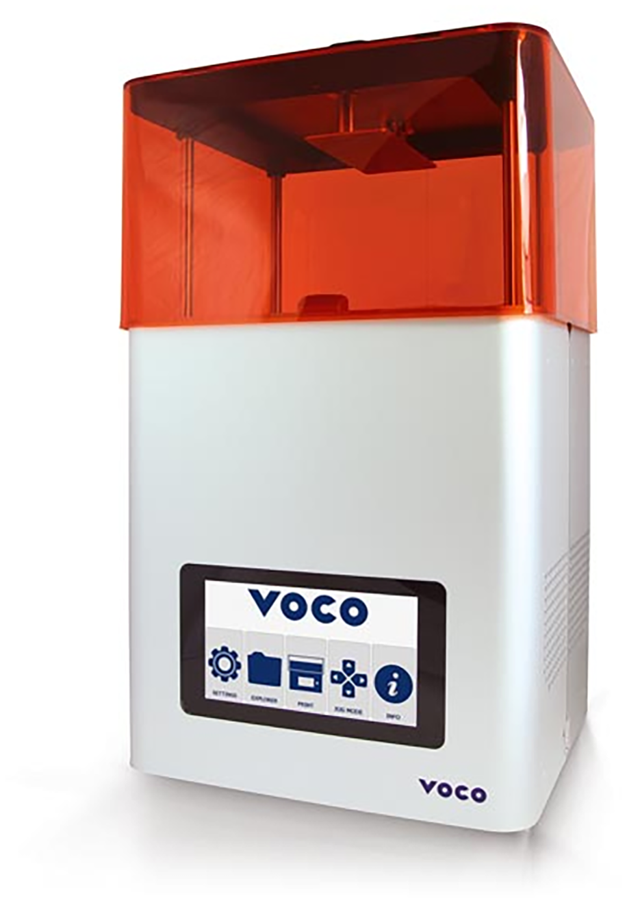
Figure 1. SolFlex 170 SLA 3D printer used to create the surgical guide [7]
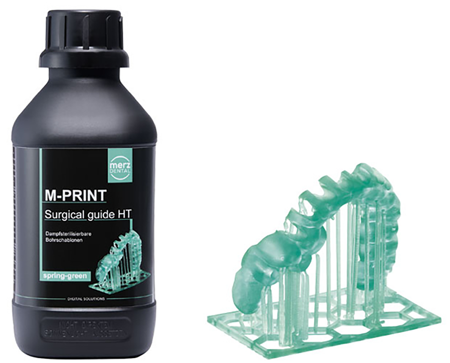
Figure 2. M-PRINT Surgical Guide HT the resin used in this study [8]
In general, the resins used in dental practice are primarily acrylic-based. Most are used in the manufacture of partial or complete dentures, and are composed of PMMA (polymethyl methacrylate) [9]. These materials are typically supplied as a two-component system: the polymer in powder form and the monomer in liquid form [10]. More often than not, the monomer component does not undergo complete polymerization, and the residual excess may cause local irritation upon contact with the soft tissues of the oral cavity. A second major category of resins consists of composite diacrylate resins. They are used primarily for direct restorations as well as for those made in the dental laboratory.
Printable resins used in dentistry must first and foremost be certified as biomaterials suitable for contact with human tissues. For the manufacturing of surgical guides, it is essential to use resin mixtures that do not cause local irritation upon contact with soft tissues and that can be disinfected and/or sterilized after manufacturing, given their direct contact with the patient’s blood. There are surgical guide resins available on the market that can be disinfected using various alcoholic and non-alcoholic solutions; however, as a precautionary measure, it is recommended to use resins that can undergo thermal sterilization after processing.
The standard version of the resin (M-Print Surgical Guide) contains urethane acrylate in its composition, which provides favorable mechanical properties [11]. In the present case, the resin used, M-PRINT Surgical Guide HT (Merz Dental), allows for heat or steam sterilization under standard conditions, using packages with sealed seams at least 8 mm wide and a minimum distance of 10 mm between the guide and the edges of the package. This characteristic ensures that no areas of pressure are exerted on the sterilized piece, thereby preventing deformation during the sterilization process [8].
M-PRINT Surgical Guide HT (Merz Dental) contains numerous chemical components, each having a specific role. The main component is diurethane dimethacrylate, which consists of a mix of isomers (UDMA). It is a long-chain-length crosslinking monomer often used in light-cure systems. In dental composites, it is used as the main oligomer. It enhances mechanical properties and wear resistance in restorative materials; after curing, it forms a glassy, hard surface, ideal for a surgical guide [12–14].
Being a polymeric material, it contains different crosslinking agents. Among them, it is ethylene dimethacrylate (EDMA), also known as ethylene glycol dimethacrylate (EGDMA). It acts as a crosslinking agent when used in polymer synthesis. Thus, it improves the mechanical properties and structural integrity of the materials when its proportion increases in the mass of the mixture. Moreover, the increase in the amount results in a decrease in the shear bond strength [15]. When used for dental composites it improves the wear resistance [16,17]. Ethylene dimethacrylate is an essential for light curing resin, being responsible for forming a three-dimensional network structure [15]. The presence of ethylene dimethacrylate in the composition of the printing resin for surgical guides such as M-PRINT Surgical Guide HT allows, compared to the standard version M-PRINT Surgical guide, for both disinfection with various chemical solutions and thermal sterilization at a temperature of 134°C for at least 3 min [8].
Another substance used as crosslinking agent is 1,4-Butanediol dimethacrylate [17]. It is widely used in printable resins used in dentistry. It is essential in the production of dental materials due to its ability to enhance mechanical strength and durability. Moreover, it enhances rigidity, chemical resistance, and polymerization efficiency. Its use in 3D printing resins contributes to obtaining highly precise printed parts.
The printing resin also contains crosslinker substances. 2-[[(butylamino)carbonyl] oxy] ethyl acrylate is one of them, commonly used as a cross-linker in polymer chemistry [18]. Actually, it is a multifunctional acrylate ester with a carbamate-linked butylamino group. The design of the molecule enables a dual reactivity. The carbamate group enhances hydrogen bonding, increasing material toughness and self-healing properties. The acrylate group takes part in the radical polymerization. In particular, for 3D printing resins, it allows for obtaining complex structures with good mechanical properties due to its ability to form cross-linked networks. Thus, it enhances the functionality of printed objects [19].
Propoxylated glycerol triacrylate, also a crosslinker substance, is a low-viscosity, aliphatic trifunctional acrylate known for its low skin irritation and high reactivity. Formulated for UV/EB curable systems, it yields films with high crosslink density and improved flexibility compared to first-generation monomers like TMPTA, while maintaining fast cure speeds.
The photoinitiator of the polymerization reaction is represented by Diphenyl (2,4,6-trimethylbenzoyl) phosphine oxide (TPO). It is an organophosphorus substance used as Norrish type I photoinitiator, unlike benzophenone that is a Norrish type II photoinitiator [20]. It has a low effect on color changes at the end of polymerization and a good solubility in most monomers compared to other photoinitiators. Negative effects are related to unreacted monomers in the case of insufficient light curing [21,22]. This aspect is eliminated in the case of printable resins, where the photopolymerization process is strictly controlled.
To enable longer-term storage of resins prior to use, polymerization inhibitor substances are incorporated into their composition. In this case, mequinol is used, an active ingredient also found in medications for patients with skin depigmentation disorders [23,24]. Here, it functions as a photopolymerization inhibitor, specifically as an ultraviolet (UV) inhibitor.
In addition to these essential components, the resin also contains colorants that enhance visibility for clinicians during the positioning of the surgical guide as well as throughout the operative stages.
3. Technological workflow to obtain surgical guide using digital technologies
The most important stages in the digital technological flow of manufacturing a maxillary expander will be presented below, starting from the aspects previously presented.
3.1. Collecting necessary data
In the first stage, the data necessary for manufacturing maxillary expander are collected, namely: panoramic radiography (orthopantomogram), lateral cephalometric radiography, virtual models of both dental arches, and most importantly, maxillary CBCT X-ray. The radiological investigations are carried out in a specialized dental radiology center. The patient’s virtual models are created by combining classical and modern techniques. Thus, classical impressions of the two arches are made using a dual-consistency condensation silicone for the maxillary arch (putty and light body), while an alginate is used for the mandibular arch (Figure 3).
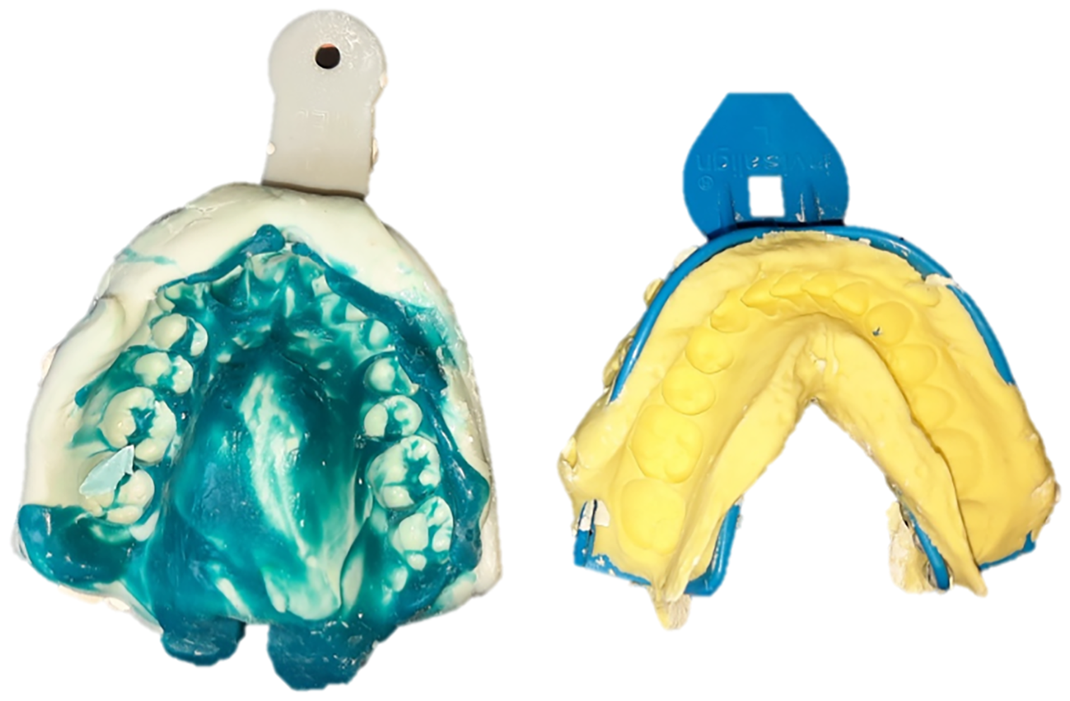
Figure 3. Impressions of the maxillary and mandibular arches
The two impressions are disinfected, following the manufacturers’ recommendations, and are subsequently used to cast the two models using type IV dental stone. The casting of the models is carried out using a vacuum mixer to eliminate air bubbles and a vibrating table so that the material could fill all the details of the impressions. After the plaster sets, the models are mounted and subsequently scanned using a laboratory scanner (Figure 4). The necessary information is thus obtained to create the virtual models required in the digital technological workflow.
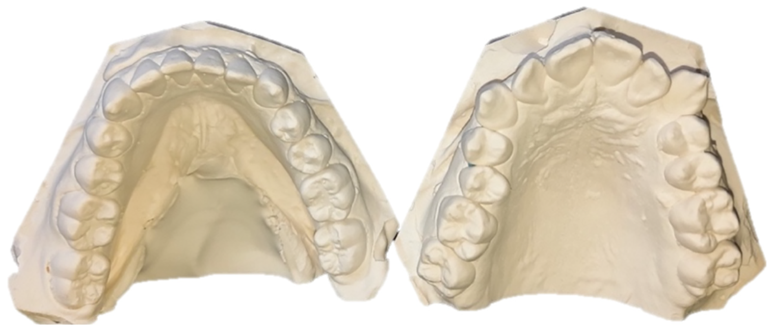
Figure 4. The appearance of the models before scanning
3.2. Overlaying the obtained data
To have a good three-dimensional image of the anatomical elements, all the previously obtained data are uploaded into the OnyxCeph3™ software [6] (Figures 5 and 6).
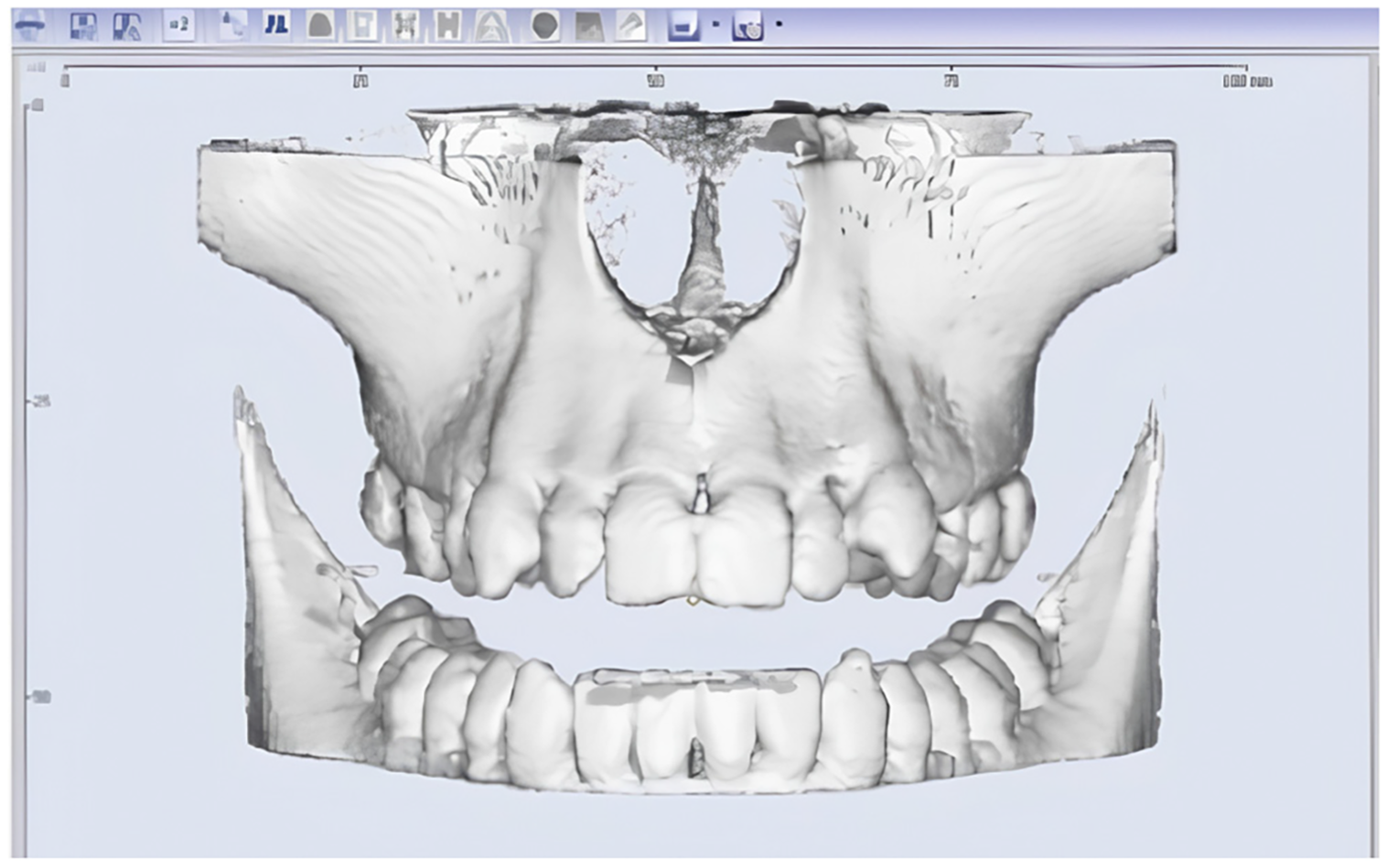
Figure 5. Three-dimensional image of the maxillary obtained by processing DICOM sections obtained from CBCT
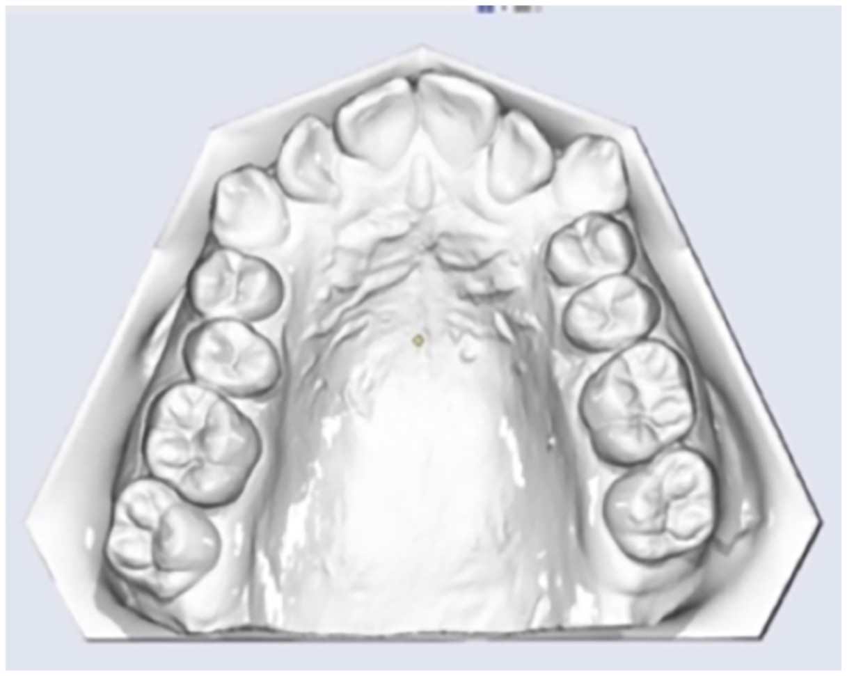
Figure 6. The virtual maxillary model obtained by scanning the functional model
Subsequently, based on clear anatomical landmarks, an alignment of all data is achieved so that a correct correlation can be established between the elements visible in the oral cavity and the supporting bone elements (Figures 7 and 8). Elements such as the middle of the incisal edge of the upper central incisor, the tip of the canine, as well as the tip of the vestibular cusps of the upper premolars and molars are chosen.
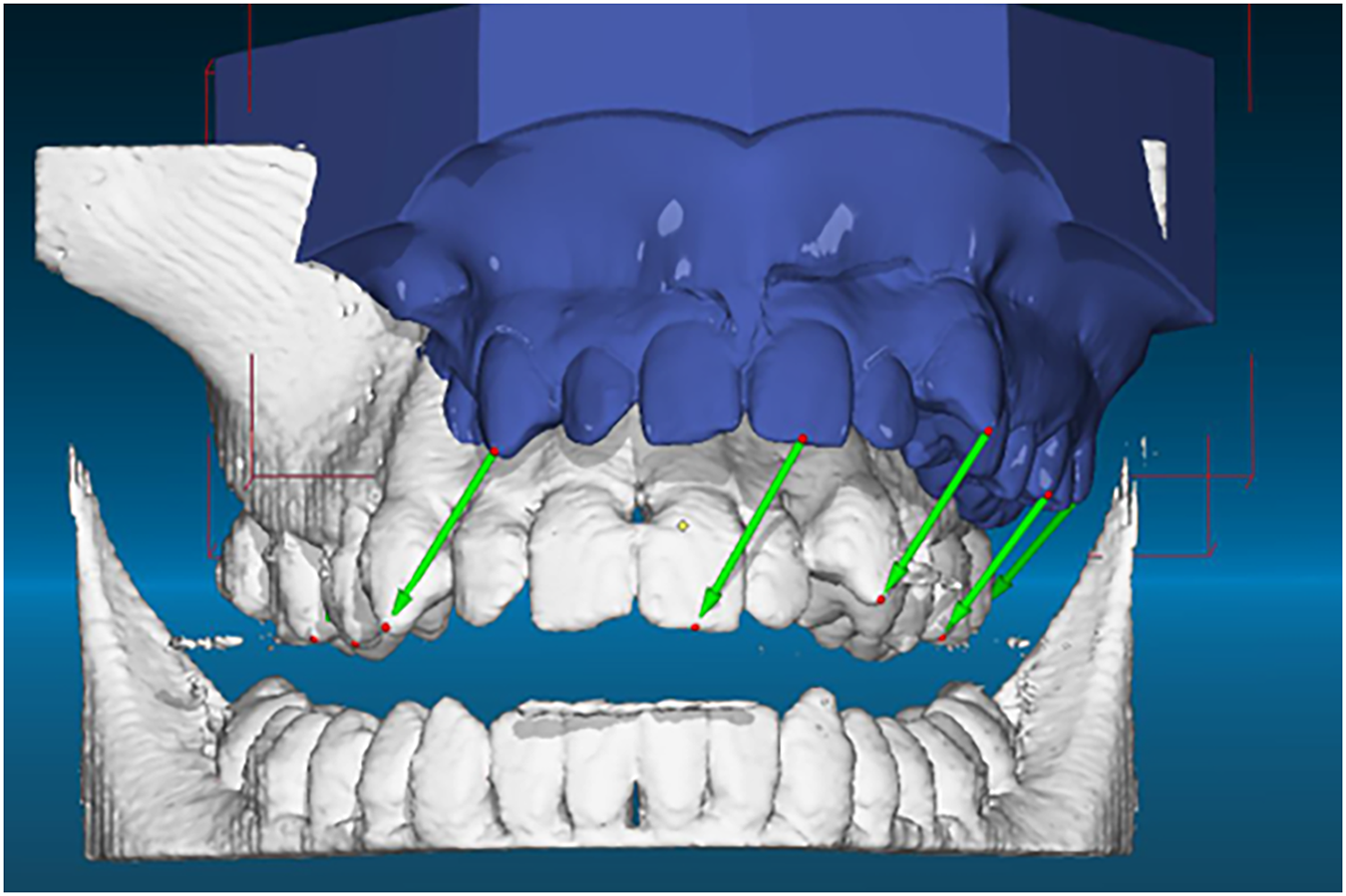
Figure 7. Selection of significant anatomical elements for aligning images obtained by CBCT and those obtained by scanning the functional model
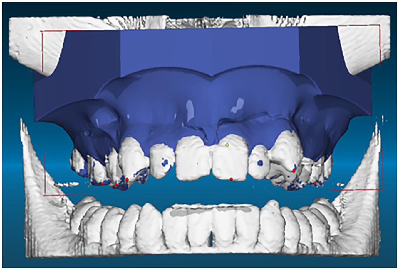
Figure 8. Appearance of the superimposed images performed by the three-dimensional analysis software
3.3. Positioning of TADs according to bone supply
Once the digital information alignment is completed, the proper type of TAD is selected from the software menu and the most appropriate location for positioning in the bone support is sought. The length of the TADs will be established depending not only on the bone supply but also on the thickness of the palatal mucosa at the level of the insertion area. The correctness of the position and the insertion axis will be verified by comparison on both lateral cephalogram and CBCT images (Figures 9 and 10). If the clinical situation does not allow the use of a single guide, it is recommended to create a surgical guide for each TAD separately.
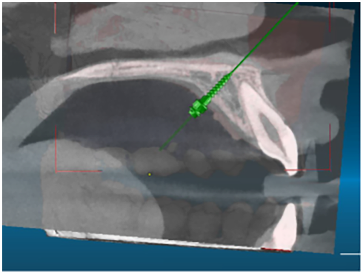
Figure 9. Checking the position and the insertion axis on the lateral cephalometric radiography
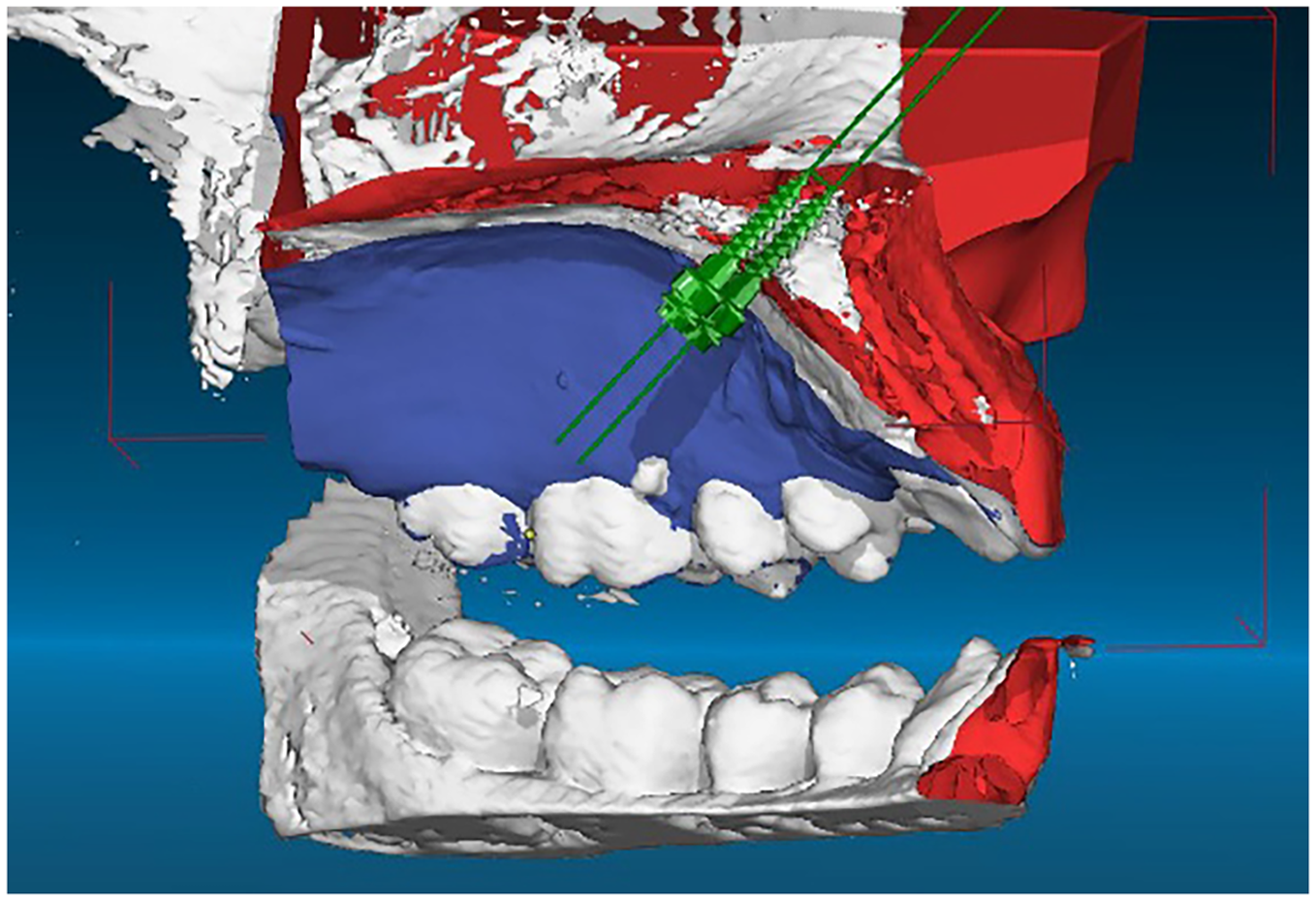
Figure 10. Checking the position and insertion axis on the superimposed three-dimensional images
3.4. Designing the virtual pattern of surgical guide
Once the anatomical landmarks, the insertion site and axis for the two TADs have been established, the design for the surgical guide is carried out. In the first phase, the teeth at which the support will be made are determined, covering not only the entire occlusal surface but also part of the vestibular face to ensure the stability of the guide. At the occlusal level, several inspection windows are created to verify the correct the guide positioning during surgical stage (Figure 11).
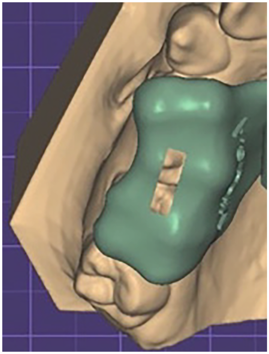
Figure 11. Design of the surgical guide at the level of the supporting teeth
The second stage is represented by the design of the slots through which the insertion of the two TADs will be carried out. To that end, the diameter of the pilot drill guide segment as well as the length chosen for the TADs will be taken into account. The guide cylinders from the surgical guide are provided with a shoulder at which both the pilot drill and the screw holder will stop, ensuring the insertion of the TAD at the desired level. Given the insertion axis, a vertical slot will be created that will allow the insertion of the pilot drill and the TAD into the surgical guide cylinder (Figures 12 and 13).
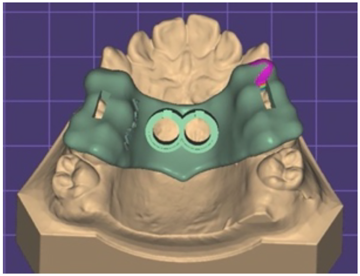
Figure 12. Diameter of guide cylinders
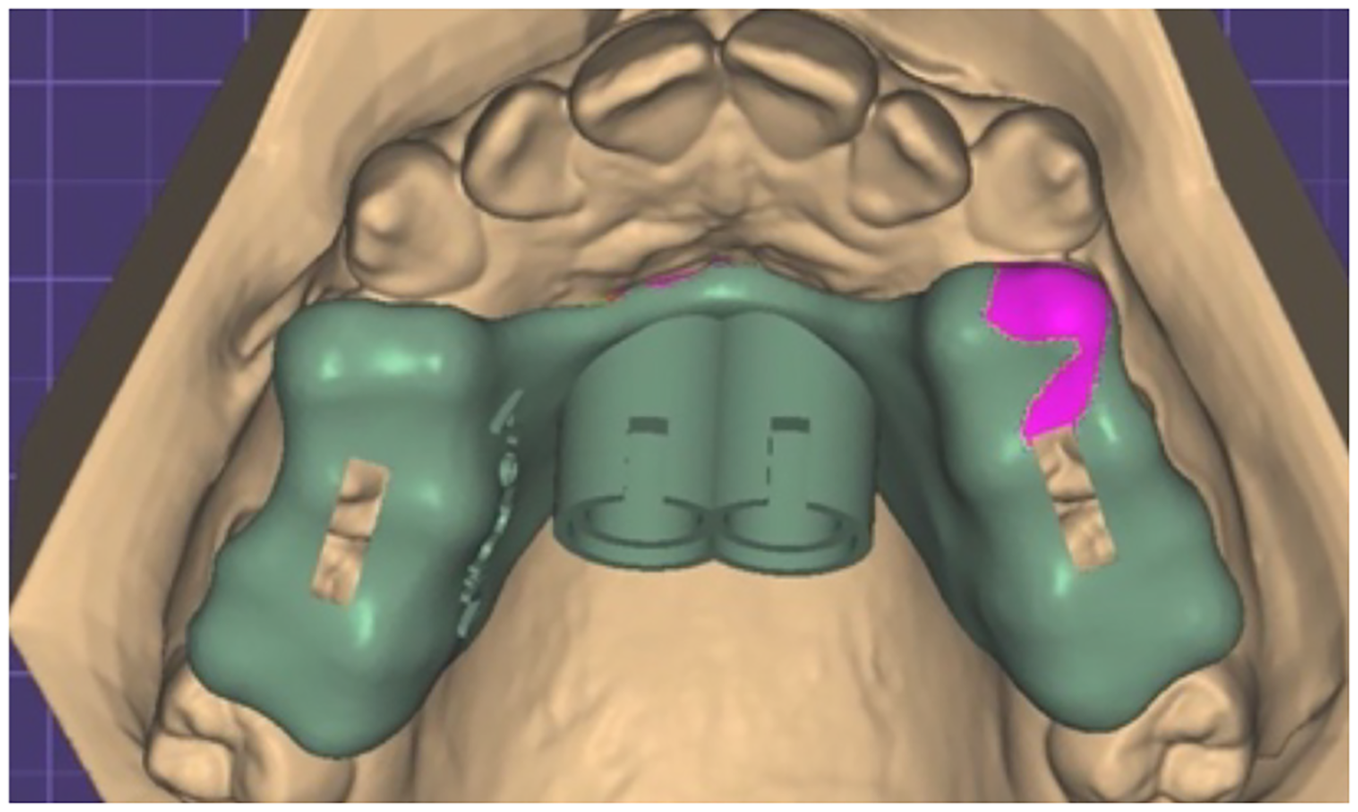
Figure 13. Appearance of the top end of the guide cylinders, the shoulder and the slot that facilitate insertion operations can be noted
The virtual pattern of the surgical guide is inspected to ensure there are no interferences with teeth or soft tissues that might create discomfort during the surgical intervention (Figure 14).

Figure 14. The visual analysis of the virtual pattern of surgical guide from all angles
3.5. Printing and processing of the surgical guide
The virtual pattern of the surgical guide is sent in stl format to the printing machine software. The dental technician establishes the three-dimensional position towards the printing platform so that the minimal interference of the support rods generated by the software can be achieved both at the level of the support areas and at the level of the insertion and guidance of the drill (Figure 15).
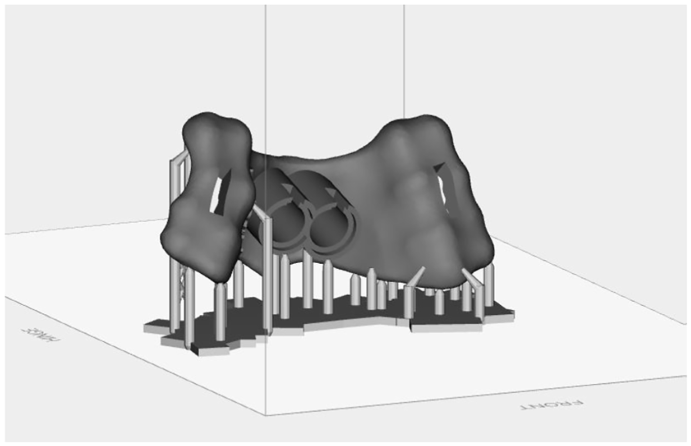
Figure 15. Positioning the virtual pattern on the printing platform
The printing accuracy is set to 50 μm (thickness of the printed layers) to minimize the degree of error. At the end of the printing process, the surgical guide is washed with isopropyl alcohol to remove liquid resin residues from the surface. In its raw form, without removing the support pins, it is introduced into the polymerization chamber for complete polymerization of the resin. The resin manufacturer recommends using the Otoflash G171 for 8 min in the post-curing process [8,25]. It ensures complete polymerization of the resin used for the surgical guide, thus preventing any complications related to incomplete polymerization [8]. Subsequently, the support pins are removed and the surfaces are finished. Before being sent to the dental office, the surgical guide is checked on the functional model obtained at the beginning of the process. The insertion of the guide, the adaptation to the occlusal surfaces (through the inspection windows), as well as the guide cylinders in relation to the screw holder are checked (Figures 16 and 17).
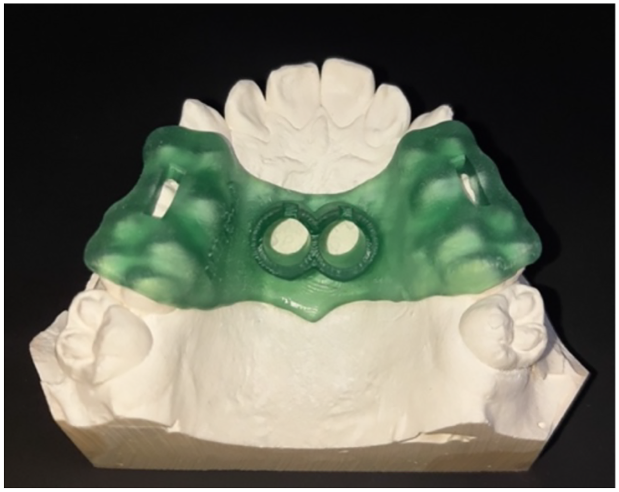
Figure 16. Checking the fitting of the surgical guide on the functional model
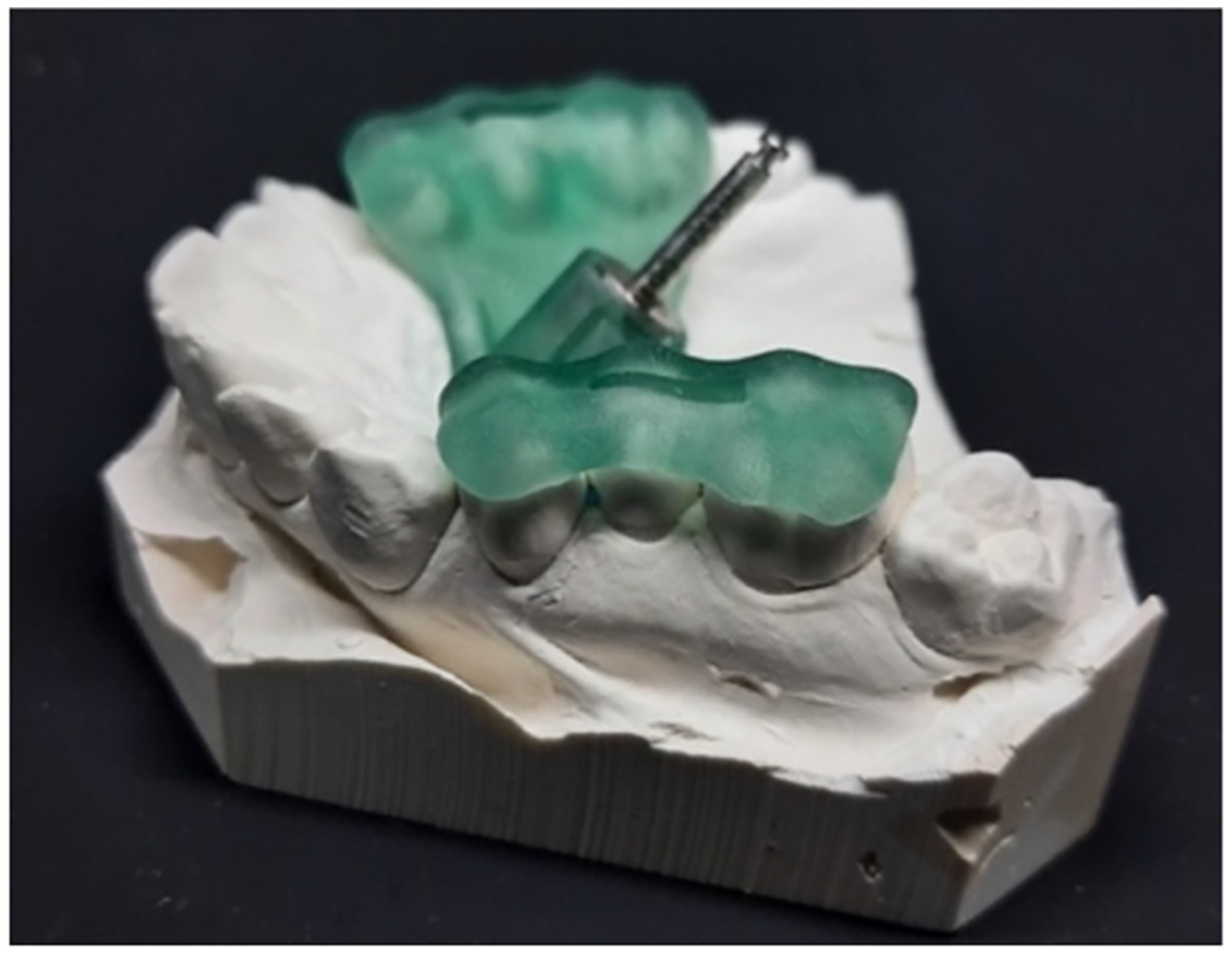
Figure 17. Checking the guide cylinders using the screw holder
The use of polymer resins in dentistry has a long history [26]. They can be presented in multiple forms, a major categorization being based on the state of polymerization. Thus, they can be polymerized resins or resins prepared for polymerization [27]. Generally, polymerized resins are found in prefabricated forms, the most well-known forms being represented by artificial acrylate teeth, celluloid strips used in direct restorations, or foils for mouth guards [28–30]. Uncured resins are used either for direct restorations in the dental office or for indirect restorations made by the dental laboratory [31,32].
In orthodontic practice, acrylic resins are mainly used for removable orthodontic appliances. However, with the development of digital technologies and the expansion of their use in orthodontics, resins have gained new uses [33]. Thus, although they do not have a direct use in orthodontic treatments, resins are used to facilitate the maneuvers and increase the chances of success [34–36]. In the case of maxillary expansion treatments, new technologies allow the manufacturing of personalized devices, in fully digital workflows, with much lower risks for dental technicians in the case of expanders made of Ni-Cr by laser sintering than by the lost-wax technique [37]. Thus, the combination of maxillary expanders and TADs has significantly improved treatment outcome [38]. As in implantology, surgical guides increase the precision of the procedures; their use for the insertion of TADs significantly reduces intraoperative accidents compared to cases where the free-hand technique is used [39–41].
The use of different devices in the technological process can also result in errors, depending on each device in part. The limitation of possible errors is partly achieved not only by frequent calibration of the devices, generally at an interval of a maximum of 14 days, but also by overlapping information coming from different sources dental scanner and CBCT. Moreover, the final product is tested on the functional model, the last step before the surgical stage. Another limitation of errors in the process of manufacturing a surgical guide for TADs is related to providing the operator with the best possible training programs, combining knowledge of anatomy, dental morphology, and material strength.
New 3D printing technologies have contributed to improving many fields of activity, including the activity of orthodontic doctors and technicians. The use of resins in clinical and technical medical flows has been made possible with the help of innovations in the field, entailing both equipment and resins composition. Thus, innovative composition and structure of resins have made them usable in surgical procedures, reducing intraoperative risks and improving the outcome of treatments.
In the case of orthodontic treatments, the introduction of surgical guides made of photopolymerizable resins has expanded the age segment of eligible patients, increased the success rate in maxillary expansion treatment, increased the precision of the surgical act, and decreased accidents and surgical complications.
Acknowledgement: Not applicable.
Funding Statement: The authors received no specific funding for this study.
Author Contributions: The authors confirm contribution to the paper as follows: study conception and design: Mihai David, Elena-Alexandra Ilie and Mihai Burlibașa; data collection: Oana Eftene, Mihaela Romanița Gligor and Mircea Popescu; analysis and interpretation of results: Radu Catalin Costea and Iuliana Babiuc; draft manuscript preparation: Viorel-Ștefan Perieanu and Mădălina Adriana Malița; material preparation and characterization: Viorel-Ștefan Perieanu and Mihai Burlibașa; critical revision of the manuscript: Viorel-Ștefan Perieanu and Mihai David. All authors reviewed and approved the final version of the manuscript.
Availability of Data and Materials: The data that support the findings of this study are available from the corresponding authors, [Viorel-Ștefan Perieanu, Mădălina Adriana Malița], upon reasonable request.
Ethics Approval: Not applicable.
Conflicts of Interest: The authors declare no conflicts of interest.
How to Cite this Article
References
- Angell EC. Treatment of irregularity of the permanent or adult teeth. Dent Cosm Mon Rec Dent Sci. 1860;1(10):540–4. doi:10.1038/sj.ebd.6400718. DOI
- Haas AJ. Rapid expansion of the maxillary dental arch and nasal cavity by opening the midpalatal suture. Angle Orthod. 1961;31(2):73–90. doi:10.1016/0278-2391(84)90207-6. DOI
- Agarwal A, Mathur R. Maxillary expansion. Int J Clin Pediatr Dent. 2010;3(3):139–46. doi:10.5005/jp-journals-10005-1069; 27616835 DOI
- Pop SI, Păcurar M, Bratu DC, Olteanu C. Aparate ortodontice. Târgu Mureș, Romania: University Press; 2018 [cited 2026 Jan 1]. Available from: https://www.librariaonline.ro/carti/aparate-ortodontice-editie-color-izabella-pop-silvia-university-press-umf-p10142981.
- Garib D, Miranda F, Palomo JM, Pugliese F, da Cunha Bastos JC, dos Santos AM, et al. Orthopedic outcomes of hybrid and conventional Hyrax expanders. Angle Orthod. 2021;91(2):178–86. doi:10.2319/060820-527.1; 33434282 DOI
- Image Instruments GmbH. OnyxCeph3™ LAB. OnyxCeph3™ Digital Orthodontics. [cited 2025 Feb 14]. Available from: https://onyxceph.eu/en/programmversionen/.
- VOCO GmbH. SolFlex 170. VOCO. [cited 2025 Feb 14]. Available from: https://www.voco.dental/en/digital/3d-print/3d-printer/solflex-170.aspx.
- Merz Dental GmbH. M-PRINT Surgical guide HT. Merz Dental. [cited 2025 Feb 14]. Available from: https://www.merz-dental.de/en/digital-solutions/3d-printing/3d-printing-liquids/m-print-surgical-guide-ht.
- Zafar MS. Prosthodontic applications of polymethyl methacrylate (PMMA): an update. Polymers. 2020;12(10):2299. doi:10.3390/polym12102299; 33049984 DOI
- Ajay R, Suma K, Ali S. Monomer modifications of denture base acrylic resin: a systematic review and meta-analysis. J Pharm Bioall Sci. 2019;11(6):112. doi:10.4103/jpbs.jpbs_34_19; 31198322 DOI
- Tzeng JJ, Yang TS, Lee WF, Chen H, Chang HM. Mechanical properties and biocompatibility of urethane acrylate-based 3D-printed denture base resin. Polymers. 2021;13(5):822. doi:10.3390/polym13050822; 33800210 DOI
- Polysciences. Diurethane Dimethacrylate (DUDMA). Polysciences, Chemistry beyond the ordinary. [cited 2025 Feb 14]. Available from: https://polysciences.com/products/diurethane-dimethacrylate-dudma.
- ARKEMA Sartomer® Americas. Technical data sheet CN1964—Urethane Methacylate. Sartomer AMERICAS. [cited 2025 Feb 14]. Available from: https://americas.sartomer.arkema.com/files/live/sites/shared_arkema/contents/pim-akeneo/f/sartomer_Urethane_US/p/MDM-13390-00-AM/TDS_CN1964_MDM-13390-00-AM_en_WW.pdf.
- Abu Bakar AA, Zainuddin MZ, Abdullah SM, Tamchek N, Mohd Noor IS, Alauddin MS, et al. The 3D printability and mechanical properties of polyhydroxybutyrate (PHB) as additives in urethane dimethacrylate (UDMA) blends polymer for medical application. Polymers. 2022;14(21):4518. doi:10.3390/polym14214518; 36365512 DOI
- Hata K, Ikeda H, Nagamatsu Y, Masaki C, Hosokawa R, Shimizu H. Development of dental poly(methyl methacrylate)-based resin for stereolithography additive manufacturing. Polymers. 2021;13(24):4435. doi:10.3390/polym13244435; 34960985 DOI
- Polysciences. Ethylene Glycol Dimethacrylate (EGDMA), ≥99.7%. Polysciences—Chemistry beyond the ordinary. [cited 2025 Feb 14]. Available from: https://polysciences.com/products/ethylene-glycol-dimethacrylate-997#:~:text=Product%20Description%20Ethylene%20Glycol%20Dimethacrylate%20(EGDMA)%20is,imparting%20mechanical%20strength%2C%20chemical%20resistance%2C%20and%20durability.
- Segerström S, Meriç G, Knarvang T, Ruyter IE. Evaluation of two matrix materials intended for fiber-reinforced polymers. Eur J Oral Sci. 2005;113(5):422–8. doi:10.1111/j.1600-0722.2005.00242.x; 16202031 DOI
- BIOSYNTH Ltd. 2-[[(Butylamino)carbonyl]oxy]ethyl acrylate. Biosynth.com. [cited 2025 Feb 14]. Available from: https://www.biosynth.com/p/FB146436/63225-53-6-2-butylaminocarbonyloxyethyl-acrylate.
- Benchchem. 2-[[(Butylamino)carbonyl]oxy]ethyl acrylate. Benchchem. [cited 2025 Feb 14]. Available from: https://www.benchchem.com/product/b1582409.
- Popescu M, Perieanu VS, Burlibașa M, Vorovenci A, Malița MA, Petri DC, et al. Comparative cost-effectiveness of resin 3D printing protocols in dental prosthodontics: a systematic review. Prosthesis. 2025;7(4):78. doi:10.3390/prosthesis7040078. DOI
- Messer-Hannemann P, Wienhold M, Esbak H, Brunner A, Schönebaum A, Schwendicke F, et al. Residual TPO content of photopolymerized additively manufactured dental occlusal splint materials. Biomedicines. 2025;13(1):44. doi:10.3390/biomedicines13010044; 39857628 DOI
- Popescu M, Malița M, Vorovenci A, Ștețiu AA, Perieanu VȘ, Costea RC, et al. Wet vs. dry dentin bonding: a systematic review and meta-analysis of adhesive performance and hybrid layer integrity. Oral. 2025;5(3):63. doi:10.3390/oral5030063. DOI
- Truswell WH. Prescription skin care products and skin rejuvenation. Facial Plast Surg Clin N Am. 2020;28(1):59–65. doi:10.1016/j.fsc.2019.09.005; 31779942 DOI
- Malița MA, Manolescu LSC, Pîrvu CF, Costea RC, Marcov EC, Burlibasa M, et al. Cumulative antibiogram: a rapid method to hinder transmission of resistant bacteria to oral cavity of newborn babies. Antibiotics. 2023;12(1):80. doi:10.3390/antibiotics12010080. DOI
- VOCO GmbH. Otoflash G171, Polymerisation unit for light-curing resin materials. VOCO. [cited 2025 Feb 14]. Available from: https://www.voco.dental/en/portaldata/1/resources/products/instructions-for-use/e1/otoflash_ifu_gb.pdf.
- Deb S. Polymers in dentistry. Proc Inst Mech Eng H. 1998;212(6):453–64. doi:10.1243/0954411981534213; 9852740 DOI
- Nanri K, Koizumi H, Hiraba H, Kikuiri T, Yoneyama T. Changes in the surface texture of pre-polymerized acrylic resin pediatric crowns following acidulated phosphate fluoride application. Dent Mater J. 2024;43(5):711–7. doi:10.4012/dmj.2024-083; 39218687 DOI
- Hao Z, Yin H, Wang L, Meng Y. Wear behavior of seven artificial resin teeth assessed with three-dimensional measurements. J Prosthet Dent. 2014;112(6):1507–12. doi:10.1016/j.prosdent.2014.04.030; 24993378 DOI
- Marcov EC, Burlibasa M, Marcov N, Costea RM, Voinescu I, Popescu M, et al. In vitro comparison of the efficiency of celluloid and metallic matrices in proximal restorations with a bulk polymer-based biomaterial. Mater Plast. 2024;61(2):1–14. doi:10.37358/mp.24.2.5715. DOI
- Marcov EC, Burlibașa M, Marcov N, Căminișteanu F, Ștețiu AA, Popescu M, et al. The evaluation of restored proximal contact areas with four direct adherent biomaterials: an in vitro study. J Funct Biomater. 2025;16(4):128. doi:10.3390/jfb16040128. DOI
- de Kuijper MCFM, Cune MS, Özcan M, Gresnigt MMM. Clinical performance of direct composite resin versus indirect restorations on endodontically treated posterior teeth: a systematic review and meta-analysis. J Prosthet Dent. 2023;130(3):295–306. doi:10.1016/j.prosdent.2021.11.009; 34980474 DOI
- Rocha MM, Carvalho AM, Coimbra FCT, de Arruda CNF, de Cássia Oliveira V, Macedo AP, et al. Complete denture hygiene solutions: antibiofilm activity and effects on physical and mechanical properties of acrylic resin. J Appl Oral Sci. 2021;29(4):e20200948. doi:10.1590/1678-7757-2020-0948; 34495105 DOI
- Netti CA, Albright MD, Jones TK. Composite resin buttons used to direct forces applied to teeth by removable orthodontic devices. J Prosthet Dent. 1992;67(5):729–31. doi:10.1016/0022-3913(92)90181-9; 1527765 DOI
- Ikenaka R, Koizumi S, Park H, Shimura M, Seimiya K, Fuchida S, et al. Accuracy of orthodontic anchor screw placement using a 3D-printed surgical guide. Cureus. 2024;16(8):e67431. doi:10.7759/cureus.67431; 39310565 DOI
- Sfarghiu GL, Oancea L, Lițescu R, Burlibașa M, Moraru L, Perieanu VS, et al. The influence of disinfectants incorporation on die stone linear expansion. Rom Biotechnol Lett. 2016;21(3):11585–90.
- Burlibașa M, Ștețiu AA, Marinescu DG, Ștețiu M, Răducanu A, Temelcea A, et al. Finite element method analysis of the stress induced upon the dental implant by the mastication process. Rom Biotechnol Lett. 2017;22(4):12706–14.
- Caministeanu F, Perieanu VS, Popa AS, Manolescu LSC, Stetiu AA, Costea RC, et al. Assessing the genotoxic impact of Ni-Cr alloys in dental prosthodontics: a preliminary comparative analysis with and without beryllium. Oral. 2025;5(2):32. doi:10.3390/oral5020032. DOI
- Pozzan L, Migliorati M, Dinelli L, Riatti R, Torelli L, Di Lenarda R, et al. Accuracy of the digital workflow for guided insertion of orthodontic palatal TADs: a step-by-step 3D analysis. Prog Orthod. 2022;23(1):27. doi:10.1186/s40510-022-00423-6. DOI
- Babiuc I, Diaconeasa MȘ, Perieanu VȘ, Malița MA, Beuran IA, Burlibașa M. Case report: digitally driven tooth autotransplantation using surgical templates and three-dimensional printed donor tooth replica. Front Oral Heal. 2025;6:1537468. doi:10.3389/froh.2025.1537468; 40177660 DOI
- Merz Dental GmbH. Printer compatibility overview. Merz Dental. [cited 2025 Feb 14]. Available from: https://www.merz-dental.de/en/digital-solutions/3d-printing/services/printer-compatibility.
- Polysciences. 1,4-Butanediol dimethacrylate, ≥92%. Polysciences—Chemistry beyond the ordinary. [cited 2025 Feb 14]. Available from: https://polysciences.com/products/14-butanediol-dimethacrylate-min-90?srsltid=AfmBOooL6Cd6T1LbUl0EfHUcVDKrnSc48E8wL-wYbnB8X-iDfpn9UgeE.