Abstract
Keywords: Biofunctional restoration; restorative dentistry; periodontal health
Over time, restorative dental materials have been primarily developed as biocompatible and inert biomaterials. Their purpose is to restore the anatomical form and functional integrity of the tooth while ensuring a durable marginal seal. These biomaterials are expected to exhibit high resistance to mechanical wear and chemical degradation in a harsh oral environment [1]. In the fabrication and clinical application, meticulous attention is given to marginal adaptation, overhanging, contour accuracy, and surface roughness. These specific factors are critical, as they can influence plaque retention, soft tissue irritation, and attachment loss. Such complications may subsequently contribute to the onset of gingivitis, periodontitis, and other periodontal pathologies [2,3].
Advancements in biomaterials science, tissue engineering, and our understanding of biological mechanisms are leading to a new hypothesis. Based on this, it can be hypothesized that restorative dental materials may evolve beyond their traditional passive role. The idea suggests the potential to develop restorative biomaterials capable of performing dual functions. These biomaterials would replace lost dental structures while also functioning as active therapeutic interfaces via timed drug release [4], modulating immune responses [5], promoting soft-tissue integration, and actively contributing to periodontal health beyond merely preventing harm. Biofunctional restorations may offer significant advantages for periodontally compromised or high-risk patients by providing active biological modulation over passive restoration, despite greater material complexity and cost.
This commentary argues that the focus of next-generation restorative dental materials should be biofunctionality. Restorative materials should promote gingival fibroblast, reduce inflammation, discourage dysbiotic biofilms, support soft tissue sealing, and ideally assist in tissue regeneration. In subsequent sections, we intend to provide a concise overview of the prevailing trends, key challenges and emerging opportunities. Based on the analysis, key design criteria for biofunctional restorative materials are defined.
2. Overview of the prevailing trends
Current research in periodontology places considerable emphasis on elucidating the biological mechanisms responsible for the initiation of gingivitis and its progression to periodontitis. Accordingly, much of the contemporary research is devoted to investigating the complex host–microbe interactions, immunomodulatory processes, microbial dysbiosis, and the underlying mechanisms of tissue regeneration. In contemporary practice, hydrogels are used for the sustained release of growth factors such as bone morphogenetic proteins (BMPs), platelet-derived growth factor (PDGF), fibroblast growth factor, and biological extracts for regenerative processes [6]. Sometimes, bioactive agents are applied topically as gels or solutions onto cleaned root surfaces during periodontal surgery to aid in modulating the local wound healing environment [7,8]. Additionally, bioactive glass-based materials are valued for their osteogenic, angiogenic, antibacterial, and immunomodulatory properties [9]. These materials are integrated into polymeric composites, hydrogels, and nanoparticles to release therapeutic ions. This ion release enhances regeneration of alveolar bone, cementum, and periodontal ligament while also modulating immune responses to foster a favorable healing environment. Ongoing research is also focused on 3D printing and bioprinting techniques to fabricate biomimetic, patient-specific scaffolds that replicate the complex architecture of periodontal tissues. These advanced approaches aim to enhance coordinated and functional tissue regeneration [10,11].
Concurrently, smart and immune-responsive restorative materials are engineered to sense and adapt to physiological stimuli. In this context, a recent study investigated antiadhesive and antibacterial dental restoratives utilizing polymeric hollow beads as carriers for active agents, demonstrating that antiadhesive composites exhibited superior biocompatibility with reduced cytotoxicity [12]. While another study demonstrated that eugenol and its derivatives, if encapsulated in polymeric nanoparticles (e.g., chitosan, zein, PLGA) within dental cements, enabled controlled and sustained release of therapeutic agents [13].
Yet another study, the investigators synthesized polymethyl methacrylate (PMMA) and graphene oxide nanocomposite with improved mechanical and antibacterial properties. Fabrication techniques ensured homogeneous dispersion and strong interaction between PMMA, fillers, and GO nanosheets [14]. While a recent study suggests that amino acid–based resin adhesive materials (ABRAMs) can promote remineralization and biofunctionality at the dentine–resin interface. They do so by stimulating apatite crystal nucleation and deposition. This process leads to the formation of a mineral layer that effectively seals the bonded dentine interface [15].
Collectively, advancements in periodontal and restorative dentistry research suggest a conceptual transformative shift from passive restorative materials to active materials endowed with dual functionality. These advanced materials not only fulfill conventional restorative requirements but also actively participate in biological signaling and tissue modulation to enhance healing and regeneration. We assume that such restorative materials can influence gingival fibroblast behavior, cytokine expression, immune modulation, and soft-tissue healing, not merely through their surface topography or marginal adaptation, but also via bioactive molecules and ions released from their surfaces.
3. Design of biofunctional restorative material
The concept of developing biofunctional restorative materials is both appealing and promising. Such materials, however, must meet rigorous criteria to be considered suitable for clinical application. From a materials science perspective, the design of biofunctional restorative materials requires a deliberate selection of material classes, architectures, and physicochemical properties to achieve both mechanical integrity and biological performance (Figure 1).
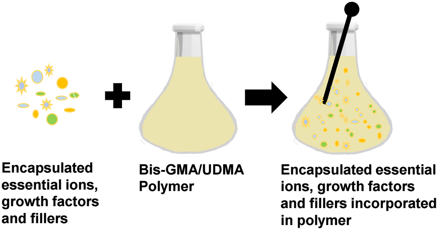
Figure 1. Schematic overview of the incorporation and functionalization of essential ions and growth factors within a resin matrix to develop a biofunctional restorative material
Firstly, controlled and sustained release of essential ions, including Ca2+, PO43−, F−, Mg2+, and Sr2, + is necessary to support fibroblast growth and tissue mineralization [16]. Secondly, the incorporation of anti-inflammatory and antioxidant agents is critical to mitigate oxidative stress within the restorative interface [17,18]. Additionally, incorporating biological extracts packed with bioactive compounds can enhance cellular proliferation in a natural way. Adding targeted growth factors such as VEGF for better blood vessel formation, BMPs for bone healing, and PDGF for tissue repair can promote regeneration. Collectively, these modifications strengthen the biofunctionality of restorative materials. However, these fillers, agents and biological extracts must be encapsulated in a polymeric material for their sustained and controlled release. Biodegradable polylactic-co-glycolic acid (PLGA) is tunable via lactide:glycolide ratios and molecular weight for weeks-to-months release, making it suitable for prolonged therapeutic delivery in periodontal tissues. However, risks such as burst release and acidic degradation by-products may exacerbate local inflammation. PMMA- and polyethylene glycol-based networks are widely investigated because of their processability, structural stability, and controllable diffusion characteristics, which support sustained release within the periodontal microenvironment [19]. Their physicochemical properties govern water uptake, diffusion kinetics, and release profiles, rendering them ideal for modulating the periodontal microenvironment [20] Nevertheless, their limited biodegradability may restrict long-term biological responsiveness. Alternatively, pH-responsive chitosan hydrogels provide inherent bioadhesion, antibacterial activity, and site-responsive release under inflammatory conditions, making them attractive for targeted periodontal delivery [21]. However, their relatively poor mechanical strength necessitates crosslinking or reinforcement. Furthermore, nanoparticle-based carriers may improve penetration and localized delivery efficiency within periodontal pockets, whereas microsphere-based systems are more suitable for prolonged depot-like release. Therefore, carrier selection should be guided by the intended therapeutic duration, inflammatory status, required mechanical stability, biodegradation behavior, and the dynamic physicochemical conditions of the oral microenvironment.
The surface properties must support soft-tissue attachment by promoting gingival fibroblast adhesion and migration, balancing roughness to prevent plaque retention yet mimicking natural extracellular matrix signals. Plasma treatment can be used to create a bioactive interface that directs favorable cellular responses while minimizing biofilm adhesion [22]. Biocompatibility with minimal cytotoxicity is crucial, especially since antimicrobial agents can inhibit fibroblast functions at certain concentrations [23].
We all know that immunomodulatory capability is vital for reducing pro-inflammatory cytokines and encouraging regenerative macrophage phenotypes, thereby avoiding chronic inflammation or fibrosis [24]. While at the same time, mechanical properties should ensure restoration strength, wear resistance, and marginal integrity to prevent crevicular leakage. Longevity under oral conditions requires resistance to enzymatic, pH, and microbial degradation, maintaining controlled release of active agents. This necessitates polymeric matrices of sufficient durability. The use of these polymers, either individually in optimized ratios or in combination (e.g., Bis-GMA/UDMA or TEGDMA/Bis-EMA), can fulfill this requirement [25]. Figure 2 schematically represents the difference between conventional passive restoration and conceptual active restoration for enhanced periodontal health.
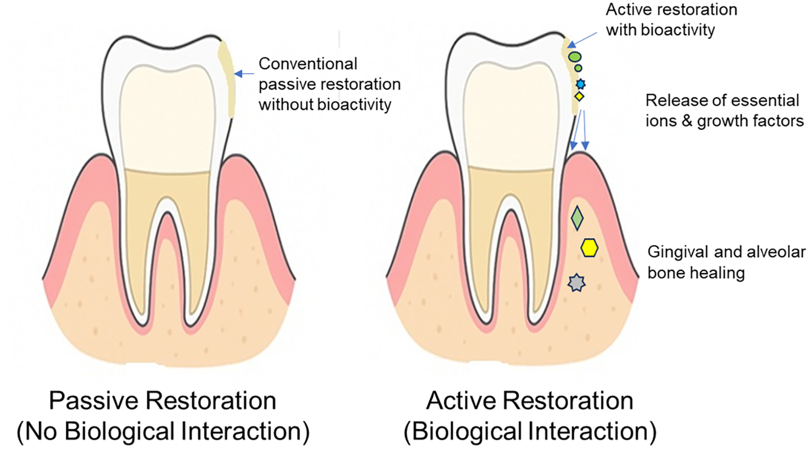
Figure 2. Schematic representation of passive restoration (left), characterized by mechanical replacement without biological interaction, and conceptual biofunctional restoration (right), illustrating active material-tissue interaction for enhanced periodontal health
4. Challenges and limitations in the development of biofunctional restoration
Developing biofunctional dental restorative materials may present several critical challenges and potential risks. Achieving a balance between antimicrobial or antiadhesive efficacy and soft-tissue compatibility remains complex. This is because certain bioactive agents may inadvertently inhibit fibroblast proliferation or interfere with collagen synthesis [26]. Therefore, restoration with precise dose optimization and controlled delivery systems within the dynamic biological environment of the periodontium is essential. Although the matrix systems discussed in the above section are considered safe, hydrolytic or enzymatic degradation over time may lead to the release of degradation by-products. This could alter local physicochemical conditions and potentially disrupt the critical balance of the oral microbiome at a susceptible periodontal site. Thus, microbiome-friendly design of biofunctional polymeric restoratives is imperative, alongside long-term evaluations to safeguard oral ecological stability from unintended therapeutic disruptions.
5. Research and implementation
The clinical translation of biofunctional restorative material demands an integrated, multidisciplinary effort. With rational material design strategies, targeted release of bioactive fillers, antioxidant/antimicrobial agents, and biological extracts is achievable. This approach can control cytotoxicity and avoid continuous localized high-dose exposure. By using pH- or enzyme-responsive polymeric matrices, on-demand release of therapeutic agents in inflammatory or dysbiotic microenvironments is possible. These strategies collectively would balance efficacy, biocompatibility, regulatory feasibility, and cost-effectiveness in developing biofunctional restorative materials. The development of advanced in vitro models with complex cell systems, biofilms, and simulated oral environments must precede in vivo validation to confirm tissue integration and the inflammatory response. Robust human clinical trials should follow, using microbiologic, gingival, and patient-centered outcomes to establish efficacy. A defined regulatory framework is crucial to ensure material safety and mechanical performance. Figure 3 presents a schematic pathway to clinical integration of biofunctional polymeric restoration for enhanced periodontal health.
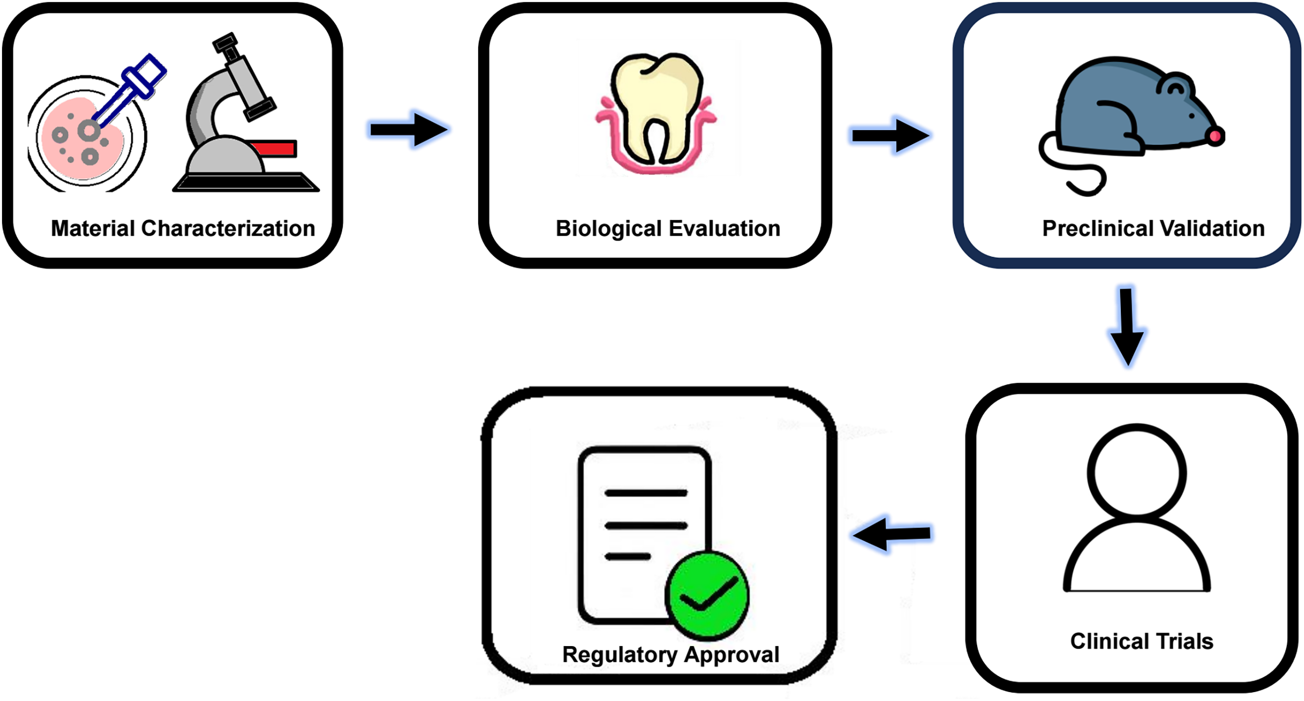
Figure 3. Schematic diagram demonstrating the pathway to clinical integration of biofunctional polymeric restoration for enhanced periodontal health
The evolving concept of biofunctionality is particularly compelling. It serves a dual role by fulfilling conventional restorative requirements while simultaneously interacting with the biological microenvironment. Through signaling and tissue modulation, it contributes to enhancing periodontal health. Realizing this concept requires the development of materials capable of controlled, safe, and biocompatible release of therapeutic agents, accompanied by measurable clinical outcomes. Progress toward this goal depends on interdisciplinary collaboration, robust standardization frameworks, and regulatory approval and translational challenges to bring this conceptual dental restorative into routine clinical practice.
Acknowledgement: Not applicable.
Funding Statement: The authors received no specific funding for this study.
Author Contributions: Aftab Ahmed Khan conceptualized the commentary, critical manuscript review and drafted the final manuscript. Mohammed Abdullah Alhadi contributed to the literature search, figure preparation support, and initial manuscript writing. Ahmed Abdullah Alhadi assisted in drafting sections on clinical implications and provided technical editing. All authors reviewed and approved the final version of the manuscript.
Availability of Data and Materials: No original data were generated or analyzed. All referenced papers are publicly available.
Ethics Approval: This commentary does not involve human or animal subjects, clinical trials, or experimental data requiring ethical approval.
Conflicts of Interest: The authors declare no conflicts of interest.
How to Cite this Article
References
- Khan AA, Zafar MS, Fareed MA, AlMufareh NA, Alshehri F, AlSunbul H, et al. Fiber-reinforced composites in dentistry-An insight into adhesion aspects of the material and the restored tooth construct. Dent Mater. 2023;39(2):141–51. doi:10.1016/j.dental.2022.12.003; 36604257 DOI
- Hajaj T, Rominu M, Constantin GD, Dobos M, Veja I. Influence of marginal tooth preparation designs on periodontal health and long-term stability: a narrative review. J Clin Med. 2025;14(19):7038. doi:10.3390/jcm14197038; 41096118 DOI
- Nafz P, Kocher T, Pink C, Baumeister SE, Reckelkamm S, Samietz S, et al. Associations between restoration margins and adjacent periodontal status—longitudinal results from SHIP-TREND. J Clin Periodontol. 2026;53(4):520–8. doi:10.1111/jcpe.70082; 41431295 DOI
- Al-Mutairi M, Al-Majed I, Khan AA, Bari A, Javed R, Al-Sadon O. Exploring the chemical and mechanical properties of functionalized bioactive glass powder-based experimental pit and fissure sealants. Polym Compos. 2025;46(8):7516–25. doi:10.1002/pc.29446. DOI
- Elazzouni DH, Alsunbul H, Almnea RA, Awadh W, Alshehri A, Alhaidry H, et al. Artificial Intelligence in the synthesis and application of advanced dental biomaterials: a narrative review of probabilities and challenges. Rev Adv Mater Sci. 2026;65(1):20250205. doi:10.1515/rams-2025-0205. DOI
- Shan BH, Wu FG. Hydrogel-based growth factor delivery platforms: strategies and recent advances. Adv Mater. 2024;36(5):e2210707. doi:10.1002/adma.202210707; 37009859 DOI
- Zhu Y, Tao C, Goh C, Shrestha A. Innovative biomaterials for the treatment of periodontal disease. Front Dent Med. 2023;4:1163562. doi:10.3389/fdmed.2023.1163562; 39916927 DOI
- Xiang C, Zhang L, Tao E. Research progress of enamel matrix derivative on periodontal tissue regeneration: a narrative review. Front Dent Med. 2025;6:1611402. doi:10.3389/fdmed.2025.1611402; 40661224 DOI
- Taye MB, Ningsih HS, Shih SJ. Exploring the advancements in surface-modified bioactive glass: enhancing antibacterial activity, promoting angiogenesis, and modulating bioactivity. J Nanopart Res. 2024;26(2):28. doi:10.1007/s11051-024-05935-2. DOI
- Zhu Y, Zhang X, Chang G, Deng S, Chan HF. Bioactive glass in tissue regeneration: unveiling recent advances in regenerative strategies and applications. Adv Mater. 2025;37(2):2312964. doi:10.1002/adma.202312964; 39014919 DOI
- Molli VLP, Pasupuleti MK, Thakkar R, Penmetsa GS, Parmar D, Jahagirdar A, et al. Advanced biomaterials in periodontal regeneration: a systematic review of systematic reviews. J Int Oral Health. 2025;17(3):174–87. doi:10.4103/jioh.jioh_259_24. DOI
- Sousa AC, Mascarenhas P, Polido M, Vasconcelos e Cruz J. Natural antibacterial compounds with potential for incorporation into dental adhesives: a systematic review. Polymers. 2024;16(22):3217. doi:10.3390/polym16223217; 39599308 DOI
- Beketova A, Tzanakakis EC, Vouvoudi E, Anastasiadis K, Rigos AE, Pandoleon P, et al. Zirconia nanoparticles as reinforcing agents for contemporary dental luting cements: physicochemical properties and shear bond strength to monolithic zirconia. Int J Mol Sci. 2023;24(3):2067. doi:10.3390/ijms24032067; 36768390 DOI
- Hassan AK, Hamidinezhad H, Al-Bermany E. Antibacterial activity and optical behavior for restoration of micro and nano dental fillers using functional graphene nanosheets with polymethyl methacrylate. Nano Biomed Eng. 2024;16(4):652–64. doi:10.26599/nbe.2024.9290075. DOI
- Sauro S, Carvalho RM, Ferracane J. The rise of advanced bioactive restorative materials: are they redefining operative dentistry? Dent Mater. 2025;41(11):1411–29. doi:10.1016/j.dental.2025.08.003; 40803933 DOI
- Yang Q, Zheng W, Zhao Y, Shi Y, Wang Y, Sun H, et al. Advancing dentin remineralization: exploring amorphous calcium phosphate and its stabilizers in biomimetic approaches. Dent Mater. 2024;40(8):1282–95. doi:10.1016/j.dental.2024.06.013; 38871525 DOI
- Zhang Y, Quindoza GM III, Mizuno HL, Nakagawa Y, Tanaka T, Anraku Y, et al. Preparation of hydroxyapatite-aligned collagen sheets and their evaluation for fibroblast adhesion and collagen secretion. ACS Biomater Sci Eng. 2025;11(2):1072–83. doi:10.1021/acsbiomaterials.4c01617; 39847580 DOI
- Farag AF, Yassin HH, Gamal AY, El Badawi N, Abdalwahab MM. Effects of different curcumin concentrations on human periodontal ligament fibroblast adhesion and proliferation on periodontally involved root surfaces: in-vitro study. J Oral Biol Craniofacial Res. 2025;15(4):729–36. doi:10.1016/j.jobcr.2025.04.006; 40352479 DOI
- Visan AI, Popescu-Pelin G, Socol G. Degradation behavior of polymers used as coating materials for drug delivery-a basic review. Polymers. 2021;13(8):1272. doi:10.3390/polym13081272; 33919820 DOI
- Luan J, Li R, Xu W, Sun H, Li Q, Wang D, et al. Functional biomaterials for comprehensive periodontitis therapy. Acta Pharm Sin B. 2023;13(6):2310–33. doi:10.1016/j.apsb.2022.10.026; 37425066 DOI
- Singh J, Nayak P. pH-responsive polymers for drug delivery: trends and opportunities. J Polym Sci. 2023;61(22):2828–50. doi:10.1002/pol.20230403. DOI
- Pal D, Neogi S. Enhancement of adhesion of polymers by plasma treatment: a critical review. Prog Adh Adhes. 2025;9:65–108. doi:10.1002/9781394315086.ch3. DOI
- Butler J, Handy RD, Upton M, Besinis A. Review of antimicrobial nanocoatings in medicine and dentistry: mechanisms of action, biocompatibility performance, safety, and benefits compared to antibiotics. ACS Nano. 2023;17(8):7064–92. doi:10.37473/dac/10.1021/acsnano.2c12488. DOI
- Caballero-Sánchez N, Alonso-Alonso S, Nagy L. Regenerative inflammation: when immune cells help to re-build tissues. FEBS J. 2024;291(8):1597–614. doi:10.1111/febs.16693; 36440547 DOI
- Khan AA, AlKhureif AA, Mohamed BA, Bautista LSJ. Enhanced mechanical properties are possible with urethane dimethacrylate-based experimental restorative dental composite. Mater Res Express. 2020;7(10):105307. doi:10.1088/2053-1591/abbf7f. DOI
- Landenberger P, Baumann L, Gerhardt-Szép S, Rüttermann S. The effect of new anti-adhesive and antibacterial dental resin filling materials on gingival fibroblasts. Dent Mater. 2021;37(9):1416–24. doi:10.1016/j.dental.2021.06.013; 34253351 DOI