Abstract
Keywords: Redox-responsive hydrogel; thioketal crosslinking; tumor microenvironment; controlled drug release; liver cancer; gelatin-based hydrogel
Liver cancer remains one of the leading causes of cancer-related mortality worldwide, with limited therapeutic options and poor long-term prognosis [1–3]. Chemotherapy is still widely used in the clinical management of liver cancer, yet its efficacy is often compromised by non-specific drug distribution, systemic toxicity, and insufficient drug accumulation at tumor sites. Doxorubicin (DOX), a commonly used chemotherapeutic agent, exhibits potent antitumor activity but is associated with severe side effects due to its lack of selectivity [4,5]. Therefore, the development of localized and microenvironment-responsive drug delivery systems is of considerable interest for improving therapeutic efficacy while minimizing off-target toxicity.
The tumor microenvironment (TME) is characterized by a series of distinct biochemical features that differ markedly from those of normal tissues [6,7]. In liver tumors, elevated levels of reactive oxygen species (ROS), high intracellular glutathione (GSH) concentrations, mildly acidic pH, and lactate accumulation are commonly observed as a consequence of aberrant metabolism and oxidative stress [8,9]. These features not only contribute to tumor progression and drug resistance but also provide endogenous stimuli that can be exploited for site-specific drug delivery. Materials capable of responding selectively to these microenvironmental cues offer an attractive strategy for achieving tumor-targeted therapy [10].
Hydrogels have emerged as promising platforms for localized drug delivery due to their high-water content, tunable mechanical properties, and structural similarity to biological tissues [11–13]. In particular, gelatin-based hydrogels have attracted extensive attention owing to their excellent biocompatibility, biodegradability, and abundance of functional groups for chemical modification [14,15]. However, conventional gelatin hydrogels often lack sufficient responsiveness to the tumor microenvironment, leading to uncontrolled drug release and limited therapeutic precision. Introducing stimulus-responsive crosslinking motifs into gelatin networks is therefore a critical step toward enhancing their functionality for cancer therapy.
Thioketal (TK) linkages are well known for their sensitivity to redox conditions, undergoing cleavage in the presence of elevated ROS and reducing agents such as GSH [16]. Incorporation of thioketal moieties into polymeric networks has been demonstrated as an effective approach to achieving redox-triggered degradation and drug release. Compared with single-stimulus-responsive systems, materials that respond to multiple redox-related cues are particularly attractive for tumor applications, as they better reflect the complex biochemical landscape of the TME. Nevertheless, the functional integration of thioketal chemistry into gelatin-based hydrogels for liver cancer–oriented microenvironment-responsive drug delivery has not yet been sufficiently clarified.
Although thioketal-based redox-responsive materials have been widely explored in hydrogel and nanomedicine systems, their application in gelatin-based matrices remains less clearly defined in terms of functional advantage. In previously reported TK-containing systems, the primary focus has largely been placed on ROS-cleavable degradation, whereas the contribution of dynamic network chemistry to gel formation and microenvironment adaptability has received less attention. In this context, the present study is not intended to claim thioketal chemistry itself as a new concept, but rather to establish a gelatin-based hydrogel platform that integrates redox-cleavable TK linkages with dynamic Schiff-base crosslinking. Compared with conventional TK-based hydrogels, the use of gelatin provides a biocompatible and highly hydrated matrix suitable for local drug delivery, while Schiff-base-mediated crosslinking enables mild gelation and network formation under relatively gentle conditions [17,18]. Relative to existing redox-responsive gelatin systems, this design introduces a more explicit redox-degradable crosslinking motif for tumor microenvironment-triggered drug release. The main advance of this work therefore lies in the rational combination of gelatin, thioketal chemistry, and dynamic covalent crosslinking into a single platform for liver cancer-oriented, microenvironment-adaptive drug delivery.
In this study, we developed a redox-responsive thioketal-crosslinked gelatin hydrogel (TK-Gel) as a microenvironment-adaptive drug delivery platform for liver cancer. The hydrogel was formed through dynamic covalent crosslinking between Gel-NH2 and thioketal-based dialdehydes, enabling mild gelation and efficient encapsulation of doxorubicin (DOX). We systematically investigated its rheological properties, swelling behavior, degradation profiles, redox responsiveness, and microenvironment-dependent drug release under normal and tumor-mimicking conditions, and further evaluated its in vitro performance using HepG2 cells. The hydrogel is formed through dynamic covalent crosslinking between Gel-NH2 and thioketal-based dialdehydes, enabling mild gelation conditions and efficient encapsulation of DOX. We systematically investigate the rheological properties, swelling behavior, degradation profiles, and redox responsiveness of the TK-Gel hydrogel under normal and tumor-mimicking conditions. Furthermore, the microenvironment-triggered drug release behavior is examined in response to variations in pH, ROS, and GSH levels. Finally, the antitumor efficacy and intracellular redox modulation of the system are evaluated using HepG2 liver cancer cells.
By integrating redox-responsive degradation with tumor-adaptive drug release, this work aims to establish a gelatin-based hydrogel platform that combines material stability, microenvironment selectivity, and enhanced therapeutic performance, providing new insights into the design of smart hydrogels for liver cancer treatment.
Gelatin (type A, from porcine skin) was used as the polymer backbone for hydrogel preparation. Thioketal-based dialdehyde (TK-dialdehyde) was synthesized according to established procedures and used as the redox-responsive crosslinker. Doxorubicin hydrochloride (DOX) was employed as the model chemotherapeutic drug. Hydrogen peroxide (H2O2), glutathione (GSH), and other analytical-grade reagents were used as received. Phosphate-buffered saline (PBS, pH 7.4) was used to simulate normal physiological conditions, while a simulated tumor microenvironment (TME) buffer (pH 6.5, supplemented with lactate) was used to mimic tumor-relevant conditions.
2.2. Preparation of TK-gel hydrogel
Gelatin was dissolved in PBS at an elevated temperature to obtain a homogeneous Gel-NH2 solution. After cooling to room temperature, TK-dialdehyde was added under gentle stirring to initiate Schiff-base crosslinking between aldehyde groups and primary amines on gelatin chains. For drug-loaded hydrogels, DOX was added to the gelatin solution prior to crosslinker addition to ensure uniform encapsulation. The mixture was allowed to react until gelation occurred, yielding the TK-Gel hydrogel. Blank hydrogels without DOX and non-thioketal-crosslinked hydrogels were prepared using the same protocol as controls.
Fourier transform infrared (FTIR) spectra of gelatin, TK-dialdehyde, and freeze-dried TK-Gel hydrogel were recorded over the range of 4000–600 cm−1 to analyze the chemical structure and confirm Schiff-base bond formation.
Rheological properties of the precursor solution and formed hydrogels were characterized using a rotational rheometer equipped with a parallel-plate geometry. Time sweep measurements were conducted at a fixed strain and frequency to monitor gelation behavior, during which the evolution of storage modulus (G′) and loss modulus (G″) was recorded. Frequency sweep tests were performed over a defined angular frequency range to evaluate viscoelastic behavior. Steady shear viscosity was measured as a function of shear rate to assess flow behavior. All measurements were carried out at controlled temperature.
2.5. Water content and swelling ratio
The equilibrium water content of the hydrogels was determined by weighing fully swollen samples and their corresponding dry weights after freeze-drying. Water content was calculated as the percentage of water relative to the swollen mass. For swelling measurements, dried hydrogels were immersed in PBS (pH 7.4) or simulated TME buffer (pH 6.5 with lactate) until equilibrium was reached. The swelling ratio (Q) was calculated as the ratio of swollen weight to dry weight.
2.6. In vitro degradation study
Hydrogel degradation was evaluated by monitoring mass loss under different conditions. Pre-weighed hydrogel samples were incubated in PBS (pH 7.4), simulated TME buffer, or PBS containing H2O2 to simulate high ROS conditions. At predetermined time points, samples were collected, washed, lyophilized, and weighed. Mass loss was calculated as the percentage decrease relative to the initial dry mass.
2.7. Redox responsiveness assay
The redox-responsive behavior of the hydrogel was assessed using a ROS-sensitive fluorescent probe. Hydrogel samples were incubated with the probe solution under controlled conditions, and fluorescence intensity was recorded at designated time intervals. Changes in normalized fluorescence intensity were used to evaluate ROS consumption associated with thioketal cleavage. Control experiments without hydrogel or with non-thioketal hydrogels were conducted for comparison.
DOX release from the hydrogels was investigated under various microenvironmental conditions, including PBS (pH 7.4), simulated TME buffer, different concentrations of H2O2, and varying GSH levels. Drug-loaded hydrogel samples were immersed in release media and incubated at physiological temperature. At predetermined time points, aliquots of the release medium were collected and replaced with fresh buffer. The amount of released DOX was quantified by UV–vis spectroscopy, and cumulative release profiles were calculated.
HepG2 human liver cancer cells were cultured in complete growth medium under standard conditions (37°C, 5% CO2). Cells were subcultured and used for experiments at appropriate confluence.
Cell viability was evaluated using the CCK-8 assay. HepG2 cells were seeded in multiwell plates and incubated with different formulations, including blank hydrogel, free DOX, DOX-loaded hydrogel, and DOX@TK-Gel under ROS conditions. After 24 h incubation, CCK-8 reagent was added, and absorbance was measured to determine relative cell viability.
2.11. Intracellular ROS measurement
Intracellular ROS levels were quantified using the DCFH-DA fluorescent probe. HepG2 cells were treated with different formulations, followed by incubation with the probe. Fluorescence intensity was measured using a microplate reader, and ROS levels were expressed as normalized fluorescence intensity relative to control groups.
The design and working mechanism of the redox-responsive thioketal-crosslinked gelatin hydrogel (TK-Gel) are illustrated in Figure 1. Gelatin chains containing primary amine groups (Gel-NH2) were crosslinked with thioketal-based dialdehydes to form a three-dimensional hydrogel network via Schiff-base reactions. During gelation, doxorubicin (DOX) was uniformly encapsulated within the polymeric matrix.

Figure 1. Schematic illustration of the redox-responsive TK-Gel hydrogel for tumor-specific drug release and FTIR spectra
As shown in Figure 1, the TK-Gel hydrogel exhibited distinct behaviors under different microenvironmental conditions. In normal physiological environments characterized by low levels of reactive oxygen species (ROS) and glutathione (GSH), the thioketal linkages remained intact, maintaining the structural integrity of the hydrogel network and effectively retaining the loaded drug. The hydrogel thus preserved a stable and compact architecture when in contact with normal tissue cells.
In contrast, under tumor-mimicking conditions representative of the liver cancer microenvironment, the hydrogel underwent pronounced structural disruption. Elevated ROS levels, increased intracellular GSH concentrations, mildly acidic pH, and lactate accumulation collectively triggered the cleavage of thioketal linkages, resulting in progressive degradation of the hydrogel network. As depicted in the right panel of Figure 1, this degradation process facilitated the release of encapsulated DOX in the tumor microenvironment. To verify the formation of Schiff-base crosslinking, FTIR spectra of gelatin, TK-dialdehyde, and the formed TK-Gel hydrogel were collected. Gelatin exhibited the characteristic amide absorption bands, while TK-dialdehyde showed a distinct aldehyde C=O absorption band. After hydrogel formation, the TK-Gel spectrum displayed a new absorption band attributable to C=N stretching, accompanied by a weakening of the aldehyde-related signal. These spectral changes support the occurrence of a Schiff-base reaction between the amino groups of gelatin and the aldehyde groups of TK-dialdehyde during hydrogel formation.
The gelation behavior of the TK-Gel hydrogel was first evaluated by time sweep rheological measurements. As shown in Figure 2A, the storage modulus (G′) increased rapidly after an initial induction period and eventually reached a stable plateau, while the loss modulus (G″) exhibited only a moderate change. The crossover and subsequent dominance of G′ over G″ indicate the formation of a solid-like hydrogel network within approximately 20–25 min.
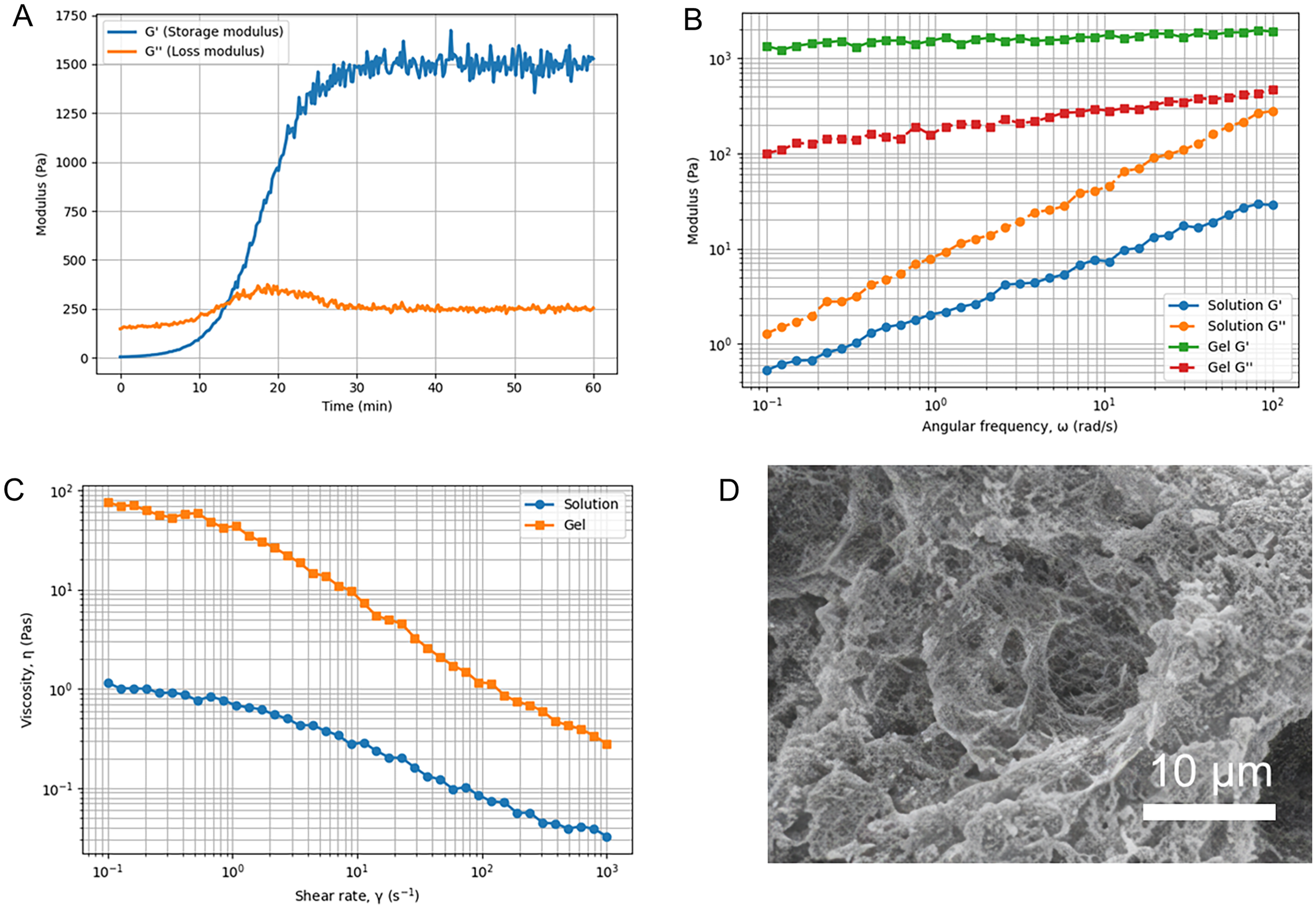
Figure 2. Rheological properties and microstructure of the TK-Gel hydrogel. (A) Time sweep measurements showing the evolution of storage modulus (G′) and loss modulus (G″) during gelation. (B) Frequency sweep results of G′ and G″ for the precursor solution and the formed hydrogel. (C) Steady shear viscosity as a function of shear rate for the solution and hydrogel. (D) Representative scanning electron microscopy (SEM) image of the freeze-dried TK-Gel hydrogel, revealing its porous and rough internal morphology
The viscoelastic properties of the hydrogel were further examined by frequency sweep measurements. As shown in Figure 2B, the precursor solution displayed a typical liquid-like behavior, with G″ exceeding G′ across the entire frequency range. In contrast, the formed TK-Gel hydrogel exhibited a markedly higher G′ than G″, with both moduli showing weak frequency dependence, characteristic of a well-developed crosslinked network.
Steady shear viscosity measurements revealed distinct flow behaviors between the solution and the hydrogel. As shown in Figure 2C, both systems exhibited shear-thinning behavior; however, the hydrogel showed significantly higher viscosity over the entire shear rate range compared to the solution, reflecting enhanced intermolecular interactions and network formation after gelation.
The internal microstructure of the TK-Gel hydrogel was visualized by SEM after freeze-drying. As shown in Figure 2D, the hydrogel exhibited a rough and irregular porous morphology with interconnected structures, indicating the formation of a three-dimensional polymer network capable of accommodating drug molecules within its matrix.
The equilibrium water content of the TK-Gel hydrogel was first evaluated in different media. As shown in Figure 3A, the hydrogel exhibited a high water content exceeding 90% in PBS (pH 7.4), confirming its highly hydrated nature. When incubated in simulated TME (pH 6.5 with lactate), a moderate but noticeable decrease in water content was observed, indicating partial network contraction under tumor-mimicking conditions.
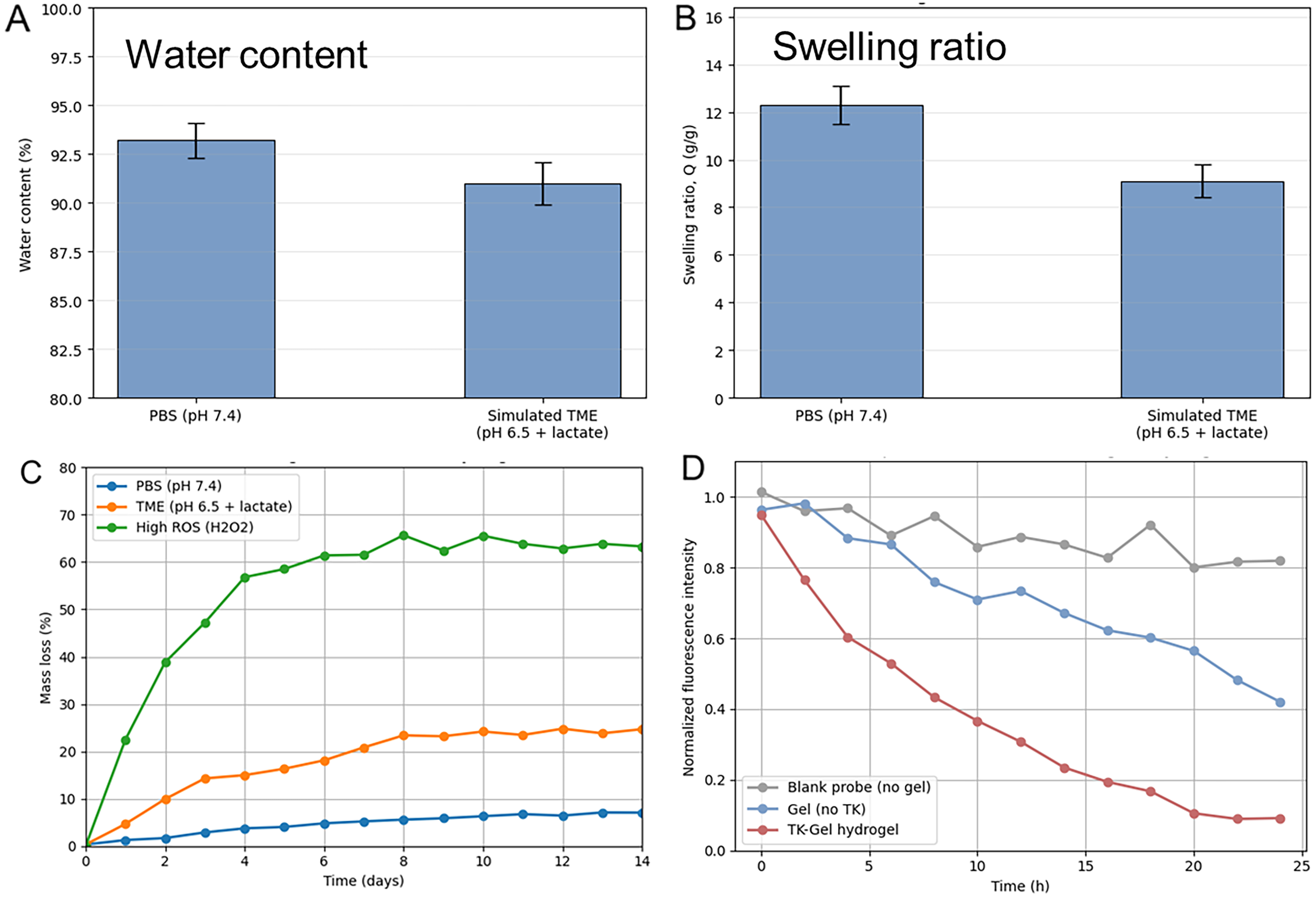
Figure 3. Swelling behavior, degradation, and redox responsiveness of the TK-Gel hydrogel. (A) Equilibrium water content of the hydrogel in PBS (pH 7.4) and simulated tumor microenvironment (TME, pH 6.5 with lactate). (B) Swelling ratio of the hydrogel under the same conditions. (C) In vitro mass loss profiles of the hydrogel incubated in PBS, simulated TME, and high ROS conditions (H2O2). (D) Time-dependent change in normalized fluorescence intensity of a ROS-sensitive probe, reflecting the redox-responsive behavior of the hydrogel
Consistent with this trend, the swelling ratio of the hydrogel was significantly higher in PBS than in simulated TME, as shown in Figure 3B. The reduced swelling ratio in the acidic and lactate-containing environment suggests enhanced network densification or reduced osmotic driving force under TME conditions.
The degradation behavior of the hydrogel was further investigated by monitoring mass loss over time. As shown in Figure 3C, the hydrogel displayed minimal mass loss in PBS over 14 days, indicating good structural stability under physiological conditions. In simulated TME, the mass loss increased gradually, reaching approximately 20–25% at day 14. In contrast, under high ROS conditions, a rapid and pronounced mass loss was observed, with more than 60% of the hydrogel mass degraded within the same period. It should be noted that the degradation behavior of the TK-Gel hydrogel cannot be attributed exclusively to thioketal cleavage, because the network also contains Schiff-base linkages that are susceptible to hydrolysis under acidic conditions. Therefore, the moderate degradation observed in simulated tumor microenvironment is likely influenced by pH-induced imine bond destabilization, whereas the substantially faster degradation under high ROS conditions more strongly indicates the contribution of thioketal bond cleavage. The present results thus support a cooperative degradation behavior governed by both acid-sensitive and redox-sensitive network components.
The redox responsiveness of the hydrogel was further confirmed by fluorescence analysis using a ROS-sensitive probe. As shown in Figure 3D, the fluorescence intensity remained relatively stable in the absence of hydrogel, while a gradual decrease was observed in the presence of a non-thioketal gel. Notably, the TK-Gel hydrogel induced a rapid and substantial decrease in fluorescence intensity over time, indicating effective ROS consumption mediated by thioketal cleavage.
Before evaluating the release behavior, the drug encapsulation performance of the TK-Gel hydrogel was quantified. The DOX-loaded hydrogel exhibited an encapsulation efficiency of 81.6 ± 2.4% and a drug loading content of 6.8 ± 0.3%, indicating that the gelatin-based network was able to effectively incorporate DOX during gel formation. Based on these results, the cumulative release profiles shown in Figure 4 were calculated relative to the initially encapsulated amount of DOX. The cumulative release behavior of DOX from the TK-Gel hydrogel was first evaluated under normal physiological and tumor-mimicking conditions. As shown in Figure 4A, DOX release in PBS (pH 7.4) proceeded slowly, reaching approximately half of the total loaded drug over 14 days. In contrast, the simulated TME condition (pH 6.5 with lactate) led to a noticeably accelerated release, particularly during the early and intermediate stages, indicating increased network permeability under tumor-like conditions.
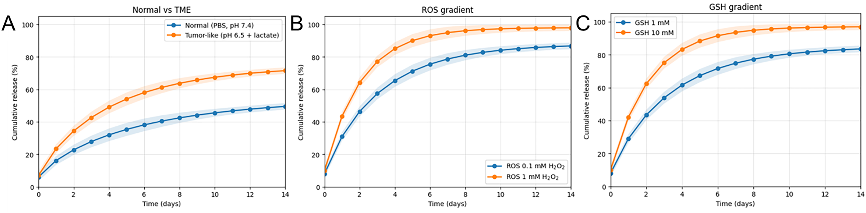
Figure 4. Microenvironment-responsive drug release behavior of the TK-Gel hydrogel. (A) DOX release in normal physiological conditions (PBS, pH 7.4) and simulated tumor microenvironment (TME, pH 6.5 with lactate). (B) DOX release under different reactive oxygen species (ROS) levels. (C) DOX release under different glutathione (GSH) concentrations
To further elucidate the role of oxidative stress, DOX release was examined under different ROS levels. As shown in Figure 4B, increasing the concentration of H2O2 resulted in a pronounced acceleration of drug release. Under high ROS conditions, the release rate was significantly enhanced during the initial days, and the cumulative release approached near-complete drug liberation by day 14. This behavior suggests that ROS directly promotes the cleavage of thioketal linkages, thereby facilitating network disassembly and drug diffusion.
The effect of reductive conditions was assessed by varying the concentration of GSH. As shown in Figure 4C, higher GSH concentrations led to faster DOX release throughout the experimental period. Compared with the 1 mM GSH condition, the 10 mM GSH environment induced a steeper release profile, indicating that reductive stimuli also contribute to the destabilization of the TK-Gel network.
The cytotoxicity of the TK-Gel hydrogel formulations toward HepG2 cells was first assessed using the CCK-8 assay. As shown in Figure 5A, untreated control cells and cells incubated with the blank hydrogel maintained high viability, indicating negligible cytotoxicity of the gelatin-based matrix. Free DOX treatment led to a marked reduction in cell viability, reflecting its strong but non-selective cytotoxic effect. In comparison, DOX-loaded hydrogel under PBS conditions exhibited higher cell viability than free DOX, suggesting a moderated drug release behavior in the absence of redox stimulation. Notably, the DOX@TK-Gel formulation under ROS conditions induced the most pronounced decrease in cell viability, indicating enhanced antitumor efficacy when the redox-responsive hydrogel was activated.
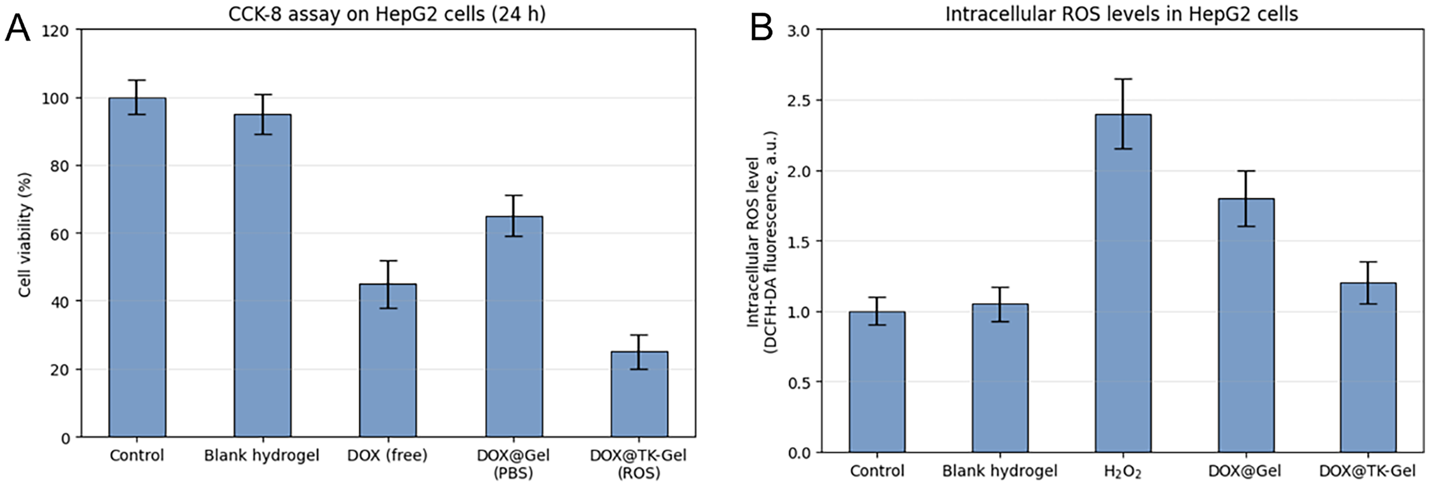
Figure 5. In vitro cytotoxicity and intracellular ROS modulation of TK-Gel in HepG2 cells. (A) Cell viability of HepG2 cells after 24 h incubation with different formulations, determined by the CCK-8 assay. (B) Intracellular reactive oxygen species (ROS) levels in HepG2 cells measured by DCFH-DA fluorescence after treatment with different formulations
Intracellular ROS levels were further quantified to investigate the redox-related cellular response. As shown in Figure 5B, control and blank hydrogel groups exhibited comparable basal ROS levels, confirming that the hydrogel matrix itself did not induce oxidative stress. Exposure to H2O2 significantly elevated intracellular ROS, serving as a positive control. Cells treated with DOX@Gel displayed increased ROS levels compared with controls, consistent with DOX-induced oxidative stress. In contrast, treatment with DOX@TK-Gel resulted in a noticeably lower ROS level than DOX@Gel, despite its higher cytotoxicity.
The schematic representation in Figure 1 highlights a rational strategy for achieving tumor-selective drug release by exploiting the redox heterogeneity between normal tissues and liver tumor microenvironments. The incorporation of thioketal linkages into the gelatin hydrogel network endows the material with intrinsic sensitivity to oxidative and reductive stimuli, which are hallmarks of malignant tissues.
In normal tissues, where ROS and GSH levels are tightly regulated, the stability of thioketal bonds ensures minimal hydrogel degradation and suppresses premature drug leakage. This feature is particularly important for reducing systemic toxicity associated with chemotherapeutic agents such as DOX. Conversely, the liver cancer microenvironment is known to exhibit elevated oxidative stress and increased intracellular reducing capacity, both of which can accelerate thioketal cleavage. The concurrent presence of high ROS and high GSH therefore provides a dual redox trigger that promotes selective hydrogel degradation at the tumor site.
Furthermore, the acidic pH and lactate-rich conditions characteristic of tumor metabolism may further destabilize the dynamic covalent network, synergistically enhancing drug release. By coupling hydrogel degradation with multiple tumor-associated biochemical cues, the TK-Gel system achieves a high level of microenvironment specificity. As conceptualized in Figure 1, this design enables localized drug liberation in tumor tissues while preserving material stability in healthy regions, thereby improving the therapeutic index and offering a promising platform for liver cancer–targeted drug delivery.
The rheological collectively confirm the successful formation of a mechanically stable TK-Gel hydrogel. The rapid increase in G′ observed during the time sweep (Figure 2A) suggests efficient crosslinking between Gel-NH2 and thioketal dialdehydes, enabling fast gelation under mild conditions. Such a gelation profile is advantageous for injectable or in situ forming hydrogel systems.
The frequency-independent plateau of G′ in Figure 2B reflects a solid-like elastic response dominated by permanent or quasi-permanent crosslinks, consistent with the formation of a chemically crosslinked network. In contrast, the liquid-like response of the precursor solution highlights the critical role of thioketal-mediated crosslinking in transforming the system from a viscous fluid into an elastic hydrogel.
The pronounced shear-thinning behavior observed in Figure 2C is beneficial for practical applications, as it facilitates handling and injection under shear while allowing rapid recovery of mechanical strength once the shear force is removed. This property is particularly relevant for localized drug delivery in irregular tumor sites.
The rough and porous morphology observed in the SEM image (Figure 2D) further supports the rheological findings. Such a heterogeneous and interconnected structure can provide sufficient space for drug encapsulation while allowing environmental stimuli to penetrate the matrix. In combination with the redox-responsive thioketal linkages, this microstructure is expected to promote environment-triggered degradation and controlled drug release in tumor tissues.
Overall, the mechanical properties and microstructural features demonstrated in Figure 2 establish a solid foundation for the subsequent investigation of redox-responsive degradation and drug release behavior of the TK-Gel hydrogel.
The results presented in Figure 3 collectively demonstrate the strong dependence of the TK-Gel hydrogel’s physical stability and degradation behavior on the surrounding microenvironment. The high water content and swelling ratio observed in PBS (Figure 3A,B) are typical characteristics of gelatin-based hydrogels and are essential for efficient molecular diffusion and drug loading. The moderate reduction in both parameters under simulated TME conditions suggests that acidic pH and lactate can subtly influence network hydration without causing abrupt structural collapse.
The degradation profiles shown in Figure 3C highlight the redox-sensitive nature of the thioketal crosslinks. While the hydrogel remains largely stable under normal physiological conditions, elevated ROS levels dramatically accelerate network breakdown. This selective degradation behavior is critical for minimizing premature drug release in healthy tissues while enabling rapid material disassembly in tumor environments characterized by oxidative stress.
Furthermore, the fluorescence results in Figure 3D provide direct evidence of ROS scavenging by the TK-Gel hydrogel. The pronounced decrease in fluorescence intensity indicates active consumption of ROS, which not only triggers thioketal cleavage but may also contribute to local modulation of oxidative stress in tumor cells. Such dual functionality—ROS-responsive degradation coupled with ROS scavenging—distinguishes the TK-Gel system from conventional hydrogels lacking redox-active linkages.
Overall, Figure 3 establishes a clear link between the tumor-associated redox microenvironment and the adaptive physicochemical behavior of the TK-Gel hydrogel, providing a mechanistic basis for its subsequent application in redox-triggered drug release and liver cancer therapy.
The drug release profiles presented in Figure 4 demonstrate that the TK-Gel hydrogel responds sensitively to multiple biochemical cues characteristic of the tumor microenvironment. The moderately enhanced release observed under simulated TME conditions (Figure 4A) can be attributed to the combined effects of acidic pH and lactate, which may weaken dynamic covalent interactions and increase polymer chain mobility. The biological relevance of GSH differs from that of acidic pH and ROS in the present delivery context. For a locally administered hydrogel, acidic and oxidative tumor-associated conditions are more likely to contribute to the initial extracellular network destabilization, whereas elevated GSH is primarily associated with the intracellular reductive environment of tumor cells. Therefore, the GSH-responsive behavior observed here is better interpreted as evidence of the broader redox adaptability of the hydrogel network and its potential responsiveness during later stages of cell-associated drug release, rather than as the dominant initial trigger of extracellular hydrogel degradation.
More pronounced differences were observed under varying redox conditions. The strong dependence of release kinetics on ROS concentration (Figure 4B) supports the design rationale of incorporating thioketal linkages as redox-cleavable crosslinks. Elevated ROS levels accelerate thioketal bond cleavage, leading to faster network degradation and more efficient drug release. This mechanism enables preferential drug liberation in oxidative tumor environments while maintaining relative stability under normal physiological conditions.
Similarly, the GSH-dependent release behavior (Figure 4C) highlights the contribution of intracellular reducing conditions to hydrogel destabilization. Tumor cells are known to maintain high GSH levels to counteract oxidative stress, and the accelerated release observed at higher GSH concentrations suggests that the TK-Gel hydrogel can exploit this reductive feature for intracellular drug delivery.
By responding to both oxidative and reductive stimuli, the TK-Gel system achieves a multi-triggered release mechanism that aligns closely with the biochemical characteristics of liver tumor tissues. This microenvironment-adaptive release behavior provides a mechanistic basis for the enhanced antitumor efficacy observed in subsequent cellular studies.
The cell viability results in Figure 5A demonstrate that incorporation of DOX into the TK-Gel hydrogel effectively alters its cytotoxic profile. While free DOX rapidly reduces cell viability, the hydrogel-based formulations enable a more controlled interaction with cells. The pronounced cytotoxicity observed for DOX@TK-Gel under ROS conditions can be attributed to redox-triggered hydrogel degradation, which promotes intracellular drug release in the oxidative tumor environment.
The intracellular ROS analysis in Figure 5B provides additional mechanistic insight. Although DOX is known to elevate intracellular ROS through redox cycling, the reduced ROS level observed in the DOX@TK-Gel group suggests that thioketal cleavage is accompanied by ROS consumption. This behavior indicates that the TK-Gel hydrogel not only serves as a drug carrier but also actively participates in redox modulation within tumor cells.
The combination of enhanced cytotoxicity and moderated intracellular ROS levels implies a distinct therapeutic mechanism for DOX@TK-Gel. Rather than relying solely on ROS overproduction for antitumor activity, the system couples redox-responsive drug release with partial ROS scavenging, which may help mitigate excessive oxidative damage to surrounding tissues. Such a balance between efficacy and redox regulation highlights the potential of TK-Gel hydrogels as microenvironment-adaptive platforms for liver cancer therapy.
A redox-responsive thioketal-crosslinked gelatin hydrogel (TK-Gel) was developed as a tumor microenvironment–adaptive drug delivery system for liver cancer therapy. The hydrogel formed a mechanically stable and highly hydrated network under mild conditions, while exhibiting pronounced microenvironment sensitivity. Swelling, degradation, and drug release behaviors were strongly dependent on tumor-relevant cues, with acidic pH, elevated reactive oxygen species, and high glutathione levels synergistically promoting thioketal cleavage and accelerated doxorubicin release, while maintaining stability under normal physiological conditions. Cellular studies using HepG2 cells demonstrated that the TK-Gel matrix was biocompatible and that redox-activated DOX@TK-Gel achieved enhanced antitumor efficacy compared with free drug and non-responsive hydrogels. In addition, thioketal cleavage was accompanied by partial intracellular ROS consumption, indicating a dual function of redox-triggered drug release and redox modulation. These results highlight the potential of TK-Gel hydrogels as microenvironment-adaptive platforms for improving the precision and safety of liver cancer drug delivery.
Acknowledgement: Not applicable.
Funding Statement: The authors received no specific funding for this study.
Author Contributions: Fajing Chen, Xiaxin Li and Yaxuan Gu contributed equally to this work, undertaking the conceptualization, methodology and original draft preparation of the manuscript. Jingjing Yi provided technical support and data interpretation for the study. Wei Cao and Lishuai Qu supervised the research, conducted formal data analysis and revised the manuscript critically. All authors reviewed and approved the final version of the manuscript.
Availability of Data and Materials: The datasets generated and/or analyzed during the current study are available from the corresponding author upon reasonable request.
Ethics Approval: Not applicable.
Conflicts of Interest: The authors declare no conflicts of interest.
Abbreviations
| ROS | Reactive oxygen species |
| TK-Gel | Thioketal-crosslinked gelatin hydrogel |
| DOX | Doxorubicin hydrochloride |
| TME | Tumor microenvironment |
| TK | Thioketal |
| GSH | High glutathione |
| H2O2 | Hydrogen peroxide |
| PBS | Phosphate-buffered saline |
| GSH | Glutathione |
How to Cite this Article
References
- Li Q, Cao M, Lei L, Yang F, Li H, Yan X, et al. Burden of liver cancer: from epidemiology to prevention. Chin J Cancer Res. 2022;34(6):554. doi:10.21147/j.issn.1000-9604.2022.06.02; 36714347 DOI
- Shiani A, Narayanan S, Pena L, Friedman M. The role of diagnosis and treatment of underlying liver disease for the prognosis of primary liver cancer. Cancer Control. 2017;24(3):1073274817729240. doi:10.1177/1073274817729240; 28975833 DOI
- Singal AG, Kanwal F, Llovet JM. Global trends in hepatocellular carcinoma epidemiology: implications for screening, prevention and therapy. Nat Rev Clin Oncol. 2023;20(12):864–84. doi:10.1038/s41571-023-00825-3; 37884736 DOI
- Carvalho C, Santos RX, Cardoso S, Correia S, Oliveira PJ, Santos MS, et al. Doxorubicin: the good, the bad and the ugly effect. Curr Med Chem. 2009;16(25):3267–85. doi:10.2174/092986709788803312; 19548866 DOI
- van der Zanden SY, Qiao X, Neefjes J. New insights into the activities and toxicities of the old anticancer drug doxorubicin. FEBS J. 2021;288(21):6095–111. doi:10.1111/febs.15583; 33022843 DOI
- Arneth B. Tumor microenvironment. Medicina. 2019;56(1):15. doi:10.3390/medicina56010015; 31906017 DOI
- Anderson NM, Simon MC. The tumor microenvironment. Curr Biol. 2020;30(16):R921–5. doi:10.1016/j.cub.2020.06.081; 32810447 DOI
- Rinaldi A, Caraffi R, Grazioli MV, Oddone N, Giardino L, Tosi G, et al. Applications of the ROS-responsive thioketal linker for the production of smart nanomedicines. Polymers. 2022;14(4):687. doi:10.3390/polym14040687; 35215600 DOI
- Rodic S, Vincent MD. Reactive oxygen species (ROS) are a key determinant of cancer’s metabolic phenotype. Int J Cancer. 2018;142(3):440–8. doi:10.1002/ijc.31069; 28940517 DOI
- Mathew J, Le T-N, Pham H-P, Kumar P, Lee C-K, Shunmugam R, et al. ROS-responsive camptothecin-linked thioketal drug delivery system based on ring-closing polymerization. Eur Polym J. 2024;202(3):112646. doi:10.1016/j.eurpolymj.2023.112646. DOI
- Xue X, Hu Y, Deng Y, Su J. Recent advances in design of functional biocompatible hydrogels for bone tissue engineering. Adv Funct Mater. 2021;31(19):2009432. doi:10.1002/adfm.202009432. DOI
- Vashist A, Vashist A, Gupta Y, Ahmad S. Recent advances in hydrogel based drug delivery systems for the human body. J Mater Chem B. 2014;2(2):147–66. doi:10.1039/c3tb21016b; 32261602 DOI
- Li J, Mooney DJ. Designing hydrogels for controlled drug delivery. Nat Rev Mater. 2016;1(12):1–17.
- Jacob S, Nair AB, Shah J, Sreeharsha N, Gupta S, Shinu P. Emerging role of hydrogels in drug delivery systems, tissue engineering and wound management. Pharmaceutics. 2021;13(3):357. doi:10.3390/pharmaceutics13030357; 33800402 DOI
- Cao H, Duan L, Zhang Y, Cao J, Zhang K. Current hydrogel advances in physicochemical and biological response-driven biomedical application diversity. Signal Transduct Target Ther. 2021;6(1):426. doi:10.1038/s41392-021-00830-x; 34916490 DOI
- Sakr MA, Sakthivel K, Hossain T, Shin SR, Siddiqua S, Kim J, et al. Recent trends in gelatin methacryloyl nanocomposite hydrogels for tissue engineering. J Biomed Mater Res Part A. 2022;110(3):708–24. doi:10.1002/jbm.a.37310; 34558808 DOI
- Andreazza R, Morales A, Pieniz S, Labidi J. Gelatin-based hydrogels: potential biomaterials for remediation. Polymers. 2023;15(4):1026. doi:10.3390/polym15041026; 36850309 DOI
- Bruce K, Marques D, Fullenkamp A, Lowell E-L, Fataki R, Ryan J, et al. Engineering of degradable linkers to improve the oxidative sensitivity of thioketal-based biomaterials. Preprint. 2025. doi:10.26434/chemrxiv-2025-90j5h. DOI